
Immune cell-mediated killing assay as shuttle session - Test your compounds in a cost-saving way
03 / 12 / 2020
Benefit from the upcoming in vitro shuttle session - scheduled in April - on our validated immune cell-mediated killing assay to assess novel immunotherapeutics in time and cost-effective manner.
The initiation of an anti-tumor immune response and further cancer cell elimination is a key step in anti-tumor immunity. One of the current promising challenges in cancer immunotherapy is the potentiation of the specific attack of tumor cells by the immune system. Interestingly, in vitro immune cell killing is recognized as perhaps the most relevant functional measure to evaluate the ability of a candidate compound to promote such an effector function of immune cells.
We designed a specialized in vitro immune cell killing assay to assess your immunomodulatory candidate - either small molecules or biologics - for their ability to promote immune cell killing activity towards tumor cells. By using live cell imaging platform, our sensitive and quantitative assay based on 96-well plate co-culture of human tumor cells (e.g. SKOV-3 ovarian, A549 or H1299 lung cancer cells…) and primary peripheral blood mononuclear cells (PBMCs) enables the kinetic monitoring and specific analysis of tumor cell death.
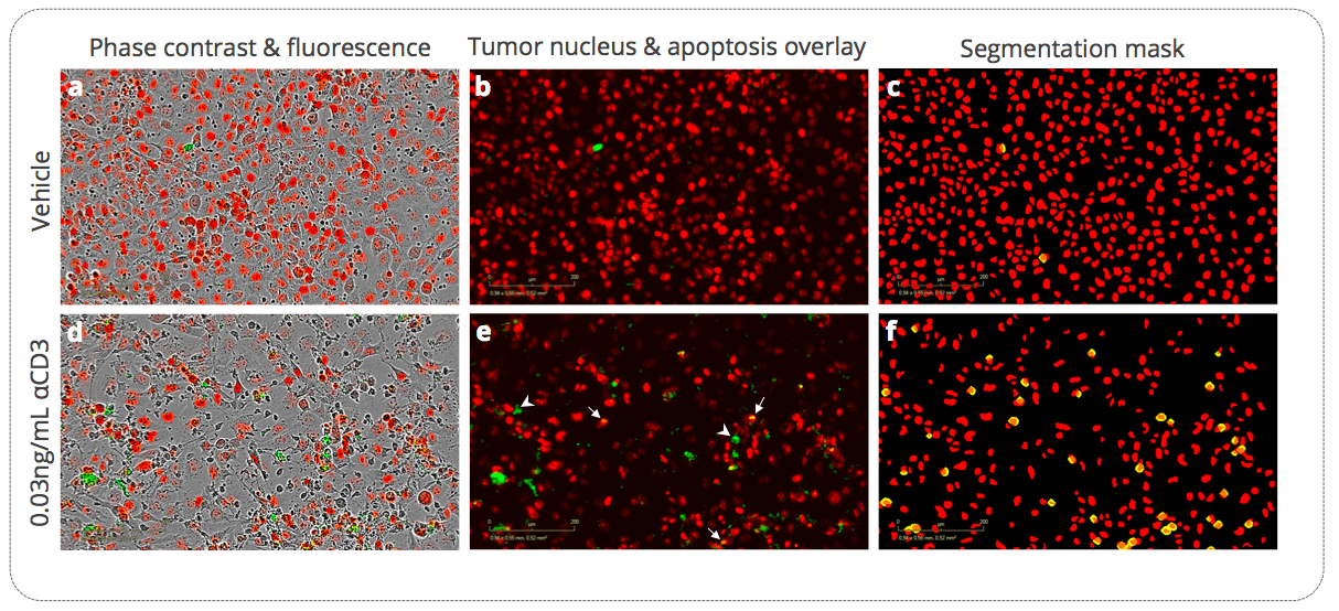
Figure 1. Immune cell-mediated killing of H1299 lung cancer cells in the presence of aCD3-activated PBMCs. H1299 tumor cells stably expressing a nuclear red fluorescent probe are cultured, at an appropriate effector:target ratio, with inactivated or aCD3-activated hPBMC. Live cell real time detection images of apoptotic H1299 tumor cells at 72h post-culture with inactivated and aCD3-activated hPBMC. Tumor cell count (nuclear red probe-expressing cells, (a, b, d, e)) and apoptosis (caspase 3/7 green fluorescent probe, (a, b, d, e)) are kinetically monitored by live cell imaging. Segmentation masks & image analysis are performed (c, f) to quantify apoptosis specifically within the tumor cell population (i.e. caspase 3/7 green fluorescent probe within the nuclear red probe-expressing cells – yellow objects). Arrows in e show apoptotic tumor cells and arrowheads apoptotic non-tumor cells.
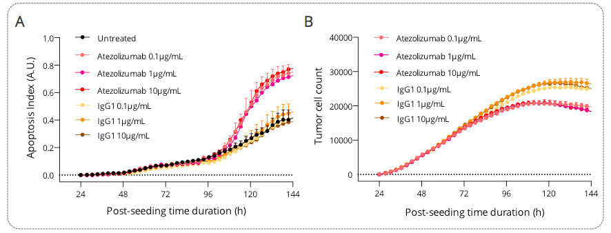
Figure 2. Real-time live cell monitoring of tumor cell killing mediated by activated PBMCs, under untreated and Atezolizumab-treated conditions. Ovarian SKOV3 tumor cells were seeded and 24h later were co-cultured with activated PBMCs, in the presence and absence of Atezolizumab or isotype control. Tumor cell apoptosis (A) and count (B) were monitored and quantified over a period of ~5 days, by mean of a caspase 3/7 fluorescent probe and a NucRed probe expression, respectively, as surrogate measures of immune cell killing activity towards tumor cells. Data were normalized and corrected to the baseline and are expressed as means ± SEM.

Visit also our Immune cell killing webpage!