-
- Model features: intact immune system, responding & non-responding models
- Tumor cell lines: Breast Cancer (4T1, EMT6), Colon Cancer (CT26, MC38), Gliobastoma (GL261), Lung Cancer (LLC1), Pancreatic cancer (Pan02), Renal Cancer (Renca), Sarcoma (MCA205)
- Tumor implantation: subcutaneous or orthotopic
- Robust treatment protocol in line with published literature data.
- Readouts: body weight, tumor size, survival
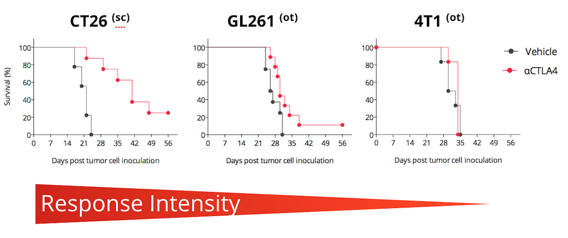
Differential CTLA4 blockade efficacy in different syngeneic mouse models CTLA4 blockade enhances survival of CT26, slightly that of GL261, but not that of 4T1 tumor-bearing mice. Mice are challenged with respective tumor cells and exposed to anti-CTLA4 antibody. In the CT26 responding model, anti-CTLA4 treatment significantly improves mouse survival. (sc, subcutaneous ; ot, orthotopic)
In vivo efficacy & mechanism of action studies for novel immunotherapies

Straightforward in vivo efficacy studies
- N=10: Standard groups of 10 mice including groups exposed to test compound alone and in combination with reference therapy.
- Weekly reports: monitoring tumor growth, body weight, and survival

Flexible sampling options
- Monitoring response over time: satellite mice, serial bleeding, intra-tumoral biopsies
- On-demand sample collection: blood, serum, plasma, tumor, organ samples

A flexible platform to quantify tumor-microenvironment & peripheral markers
- Multiplex immunophenotyping by flow cytometry & digital pathology
- Spatial transcriptomics & proteomics
Immune profiling of CT26 tumor-bearing model to anti-CTLA4 therapy
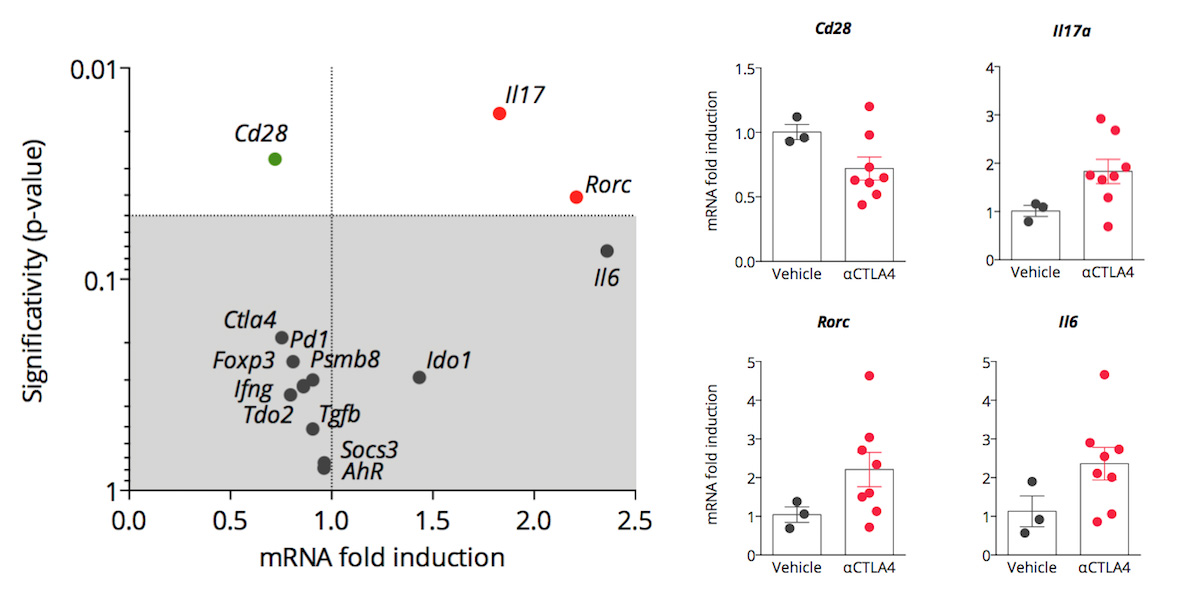
RT-qPCR analysis of key immune markers encoding genes in CT26 tumor-bearing model of anti-CTLA4 therapy. CTLA4 blockade modulates Th17-related encoding genes in TDLNs, including Rorc, Il17a and Il6.
Higher immune cell infiltration upon anti-CTLA4 therapy
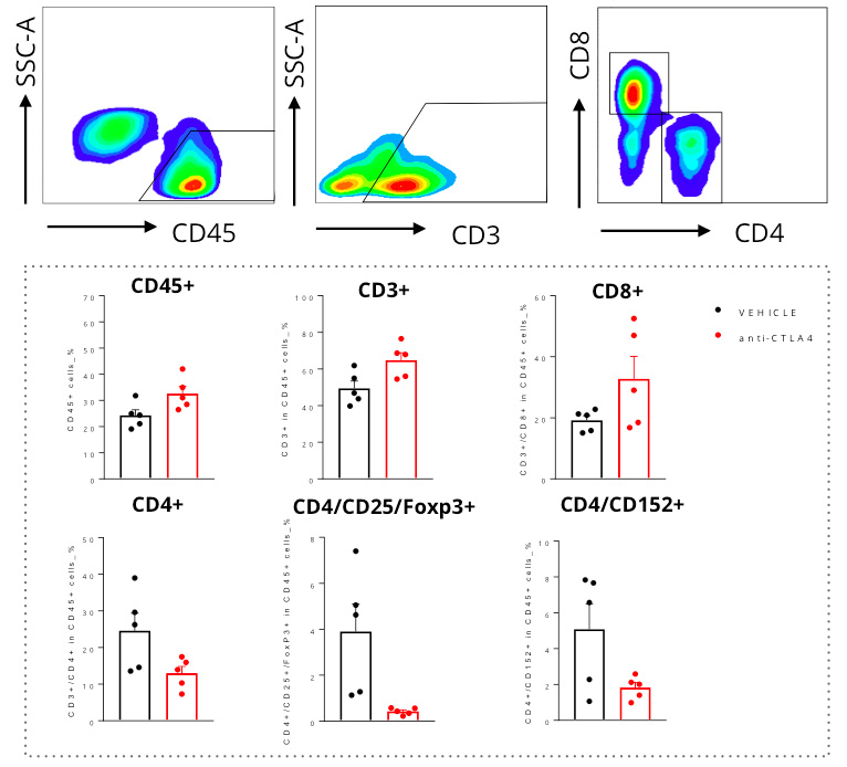
Flow cytometry analysis of CT26 tumors highlights a higher immune cell infiltration within the tumor upon CTLA4 blockade. Anti-CTLA4 antibody treatment of CT26-tumor bearing mice leads to a higher number of Tumor infiltrating lymphocytes (CD45+). This feature is associated with an increase in T cells (CD3+) and particularly with an accumulation of effector T cells (CD8+). In contrast, CD4+ T cells infiltration of the tumor is decreased upon treatment.
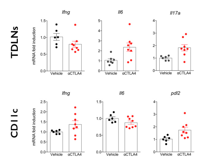
Analysis of key immune markers encoding genes by RT-qPCR.
TDLN (Tumor Draining Lymph Nodes) and CD11c + dendritic cells from CT26 tumor bearing mice exposed to anti-CTLA4 antibody or only exposed to vehicle were analyzed by RT-qPCR for specific immune markers. Results show that anti-CTLA4 exposure induces an up-regulation of Il6 and Il17 in TDLN while an icrease in Ifng and Pdl2 is observed in dendritic cells.
Why working with Explicyte?
Experts
in Immuno-Oncology
- 150+ in vivo campaigns conducted over the past 10 years
- 20+ peer-reviewed publications in key immuno-oncology journals
- Bespoke study designs based on client objectives and literature
Personalized
approach
- A dedicated study director (PhD level) from experimental plan to final report
- Weekly reports to provide regular updates & adapt experimental strategy
- Comprehensive analytical platform to decipher anti-tumor response
Your contacts

Talk to our team ! Your key contacts:
Study directors: Paul Marteau, PharmD (not on picture), Jean-Philippe Guégan, PhD Leadership: Imane Nafia, PhD (CSO), Alban Bessede, PhD (founder, CEO), Loic Cerf, MSc (COO)
Tell us about your project !
In Vivo Models Of CTLA-4 Blockade I Immuno-Oncology CRO services
As monotherapy, CTLA-4 immune checkpoint blockade installs a long-lasting anti-tumor immunity in a small fraction of patients. To assess new drugs for their capacity to improve the benefit of CTLA-4 modulators, we offer syngeneic tumor-bearing mouse models with well-characterized responses to an anti-CTLA-4 monoclonal antibody. Our models include both CTLA-4 inhibition responding and non-responding models. They enable the evaluation of novel compounds capable of enhancing the efficacy of anti-CTLA-4 antibodies and the study of predictive biomarkers to select anti-CTLA-4 responders.

