The underlying question: Does your compound exhibit immunomodulatory properties of cell migration, either by promoting the migration of effector immune cell migration, or by inhibiting the migration of cancer cells or suppressive immune cells?
- Format: 96-well plates
- Models: primary or immortalized immune cells (T cells, neutrophils, other specific subset)
- Readouts: cell migration in response to a chemoattractant – chemotaxis
- Chemoattractants: SDF1α (T-Cells), fMLP, IL-8, CXCL1 (Neutrophils), any other of interest on request
- On request: flow cytometry immunophenotyping , assessment of cytokine release, proteomics, transcriptomics
Real-time visualization of chemotactic migration of immortalized T cells toward SDF1α in a 96-well cell migration plate. Transmembrane migration of T cells was quantified using the IncuCyte® live cell analysis system.
Assay principle
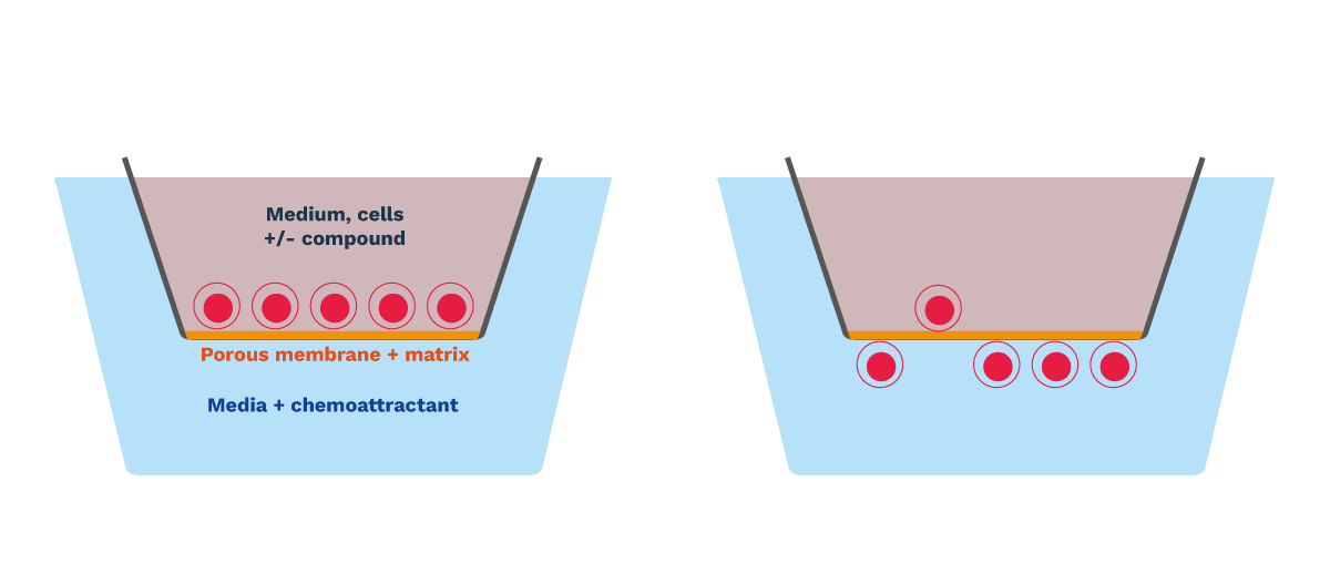
- Immune cells (T lymphocytes, neutrophils), or cancer cells (non-adherent) are seeded onto a pore-based insert, coated with extracellular matrix as needed. This upper chamber is immersed in a lower chamber, filled with culture media complemented with a chemoattractant
- Challenge of cells with drug candidates in the upper chamber, and with chemoattractants in the lower chamber
- Chemotactic cell migration from upper to lower chamber is quantified by real-time imaging over 6 to 48h
- Additional readouts: flow cytometry immunophenotyping, cytokine release, gene expression, proteomics

Testing chemotactic primary human T cell migration
Chemotactic primary T cell migration is dose-dependently induced by SDF1a over a 24h period (A). This chemoattractive effect is reversed in the presence of a CXCR4 inhibitor – AMD-3100 (B).

Human neutrophil chemotaxis assay to screen immunomodulatory activities of drug candidates
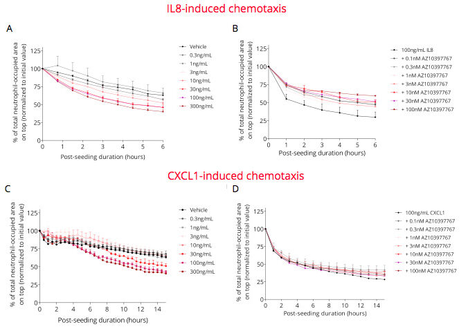
Freshly isolated human neutrophils are plated on top side of the pore-based insert, and then exposed to IL8 (A) or CXCL1 (C), in the presence and absence of a CXCR2 antagonist AZD10397767 (B, D). Cell migration monitored by real-time imaging is then analyzed over a 6h and 15h period, respectively. Chemotactic neutrophil migration is dose-dependently induced by IL8 (A) and CXCL1 (C). The CXCR2 antagonist AZD10397767 partially abolishes these IL8-induced (B) and CXCL1-induced (D) chemotactic effects.

Why working with Explicyte?
Experts
in Immuno-Oncology
- 100+ in vitro campaigns conducted over the past 10 years
- 30+ peer-reviewed publications in key immuno-oncology journals
- A comprehensive technology platform to decipher mechanism of action (activation markers, signaling pathways…)
Personalized
approach
- Isolation of specific immune cells from primary PBMCs
- Targeted discussion based on your request to design bespoke strategy & fit-for-purpose study proposal
- A dedicated study director (PhD level) from experimental plan to final report discussion
Your contacts

Talk to our team !
Paul Marteau, PharmD (preclinical study director), Imane Nafia, PhD (CSO), Loïc Cerf, MSc (COO), Alban Bessede, PhD (founder, CEO), Jean-Philippe Guégan, PhD (CTO)
Tell us about your project !
Immune Cell Migration Assays for Cancer Immunotherapies
Cell migration is a key critical process of live cells involved in many biological processes – physiological and pathological – including normal development, immune response, and many pathological processes e.g. cancer metastasis, tumor immune infiltration, inflammation, wound healing… The study of cell migration in cancer research is of particular interest as two of the main challenges in the development of novel therapeutic strategies are i) the inhibition of metastatic progression of cancer cells and ii) stimulation of the recruitment of immune cells and their infiltration into the tumor masses. Indeed, while for cancer spread throughout the body cancer cells must migrate and invade through extracellular matrix, and intravasate/extravasate, chemotactic immune cell recruitment and infiltration into the tumor (e.g. neutrophils, effector T cells, regulatory T cells (Treg), Th17, macrophages, dendritic cells) is primordial for effective anti-tumor immune response to counter disease progression. In this respect, Explicyte is offering valuable in vitro cell-based 96-w plate assays to evaluate compounds effects (with potential chemoattractive or chemorepulsive properties) on the migration of immune or cancer cells, respectively. Based on live cell time-lapse imaging, our assays unlike conventional single time-point chemotaxis/migration assays allow for full time course chemotaxis/cell migration profiles.


