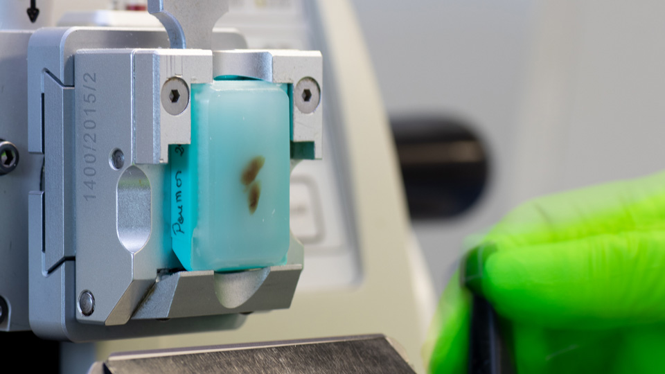
Tissue sourcing & processing
- Custom sourcing of tumor specimens through accredited biobanks
- Fixation & paraffin embedding (FFPE tissues) or tissue freezing
- Slicing of FFPE samples or cryosections, and slide preparation
- H&E coloration & annotation by trained pathologists
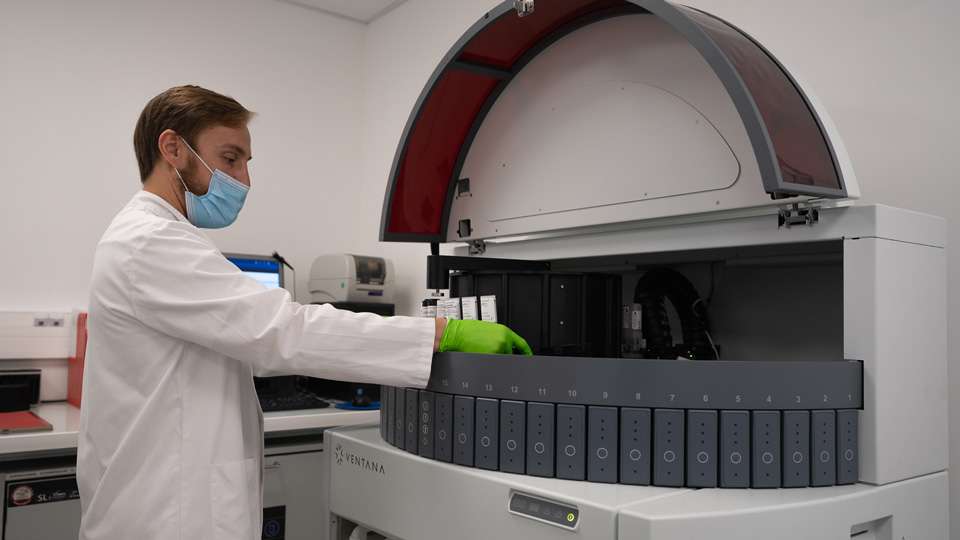
Automated IHC/IHF staining
- 2x automated Ventana Discovery XTTM systems
- Up to 200 samples stained per week including Tissue MicroArrays (TMA)
- A range of validated panels for immuno-oncology or custom panel development
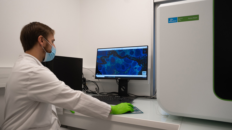
Multiplex Pathology Imaging
- Multispectral imaging with PhenoImagerTM (Akoya Biosciences, 2 systems)
- Up to 9 colors
- >100 slides acquired per week
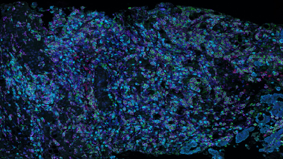
Image analysis
- Pathologist-assisted digital image analysis
- In-house capacities for AI-based tissue & cell segmentation
- A dedicated study director to tailor the analysis plan & generate biologically relevant data
Get our list of validated markers & panels (IHC/IHF) for immuno-oncology
Experts
in Immuno-Oncology
- Automated workflow for large-scale campaigns
- 150+ validated markers to assess tumor microenvironment (TME) features
- 30+ peer-reviewed publications in key immuno-oncology journals
Personalized
approach
- Custom markers developed and validated as monoplex or within multiplexed panels
- A dedicated study director (PhD) to tailor the experimental plan to your need and discuss results
- Complementary technologies to generate string dataset: spatial transcriptiomics, single-cell RNA Seq, proteomics, flow cytometry, …
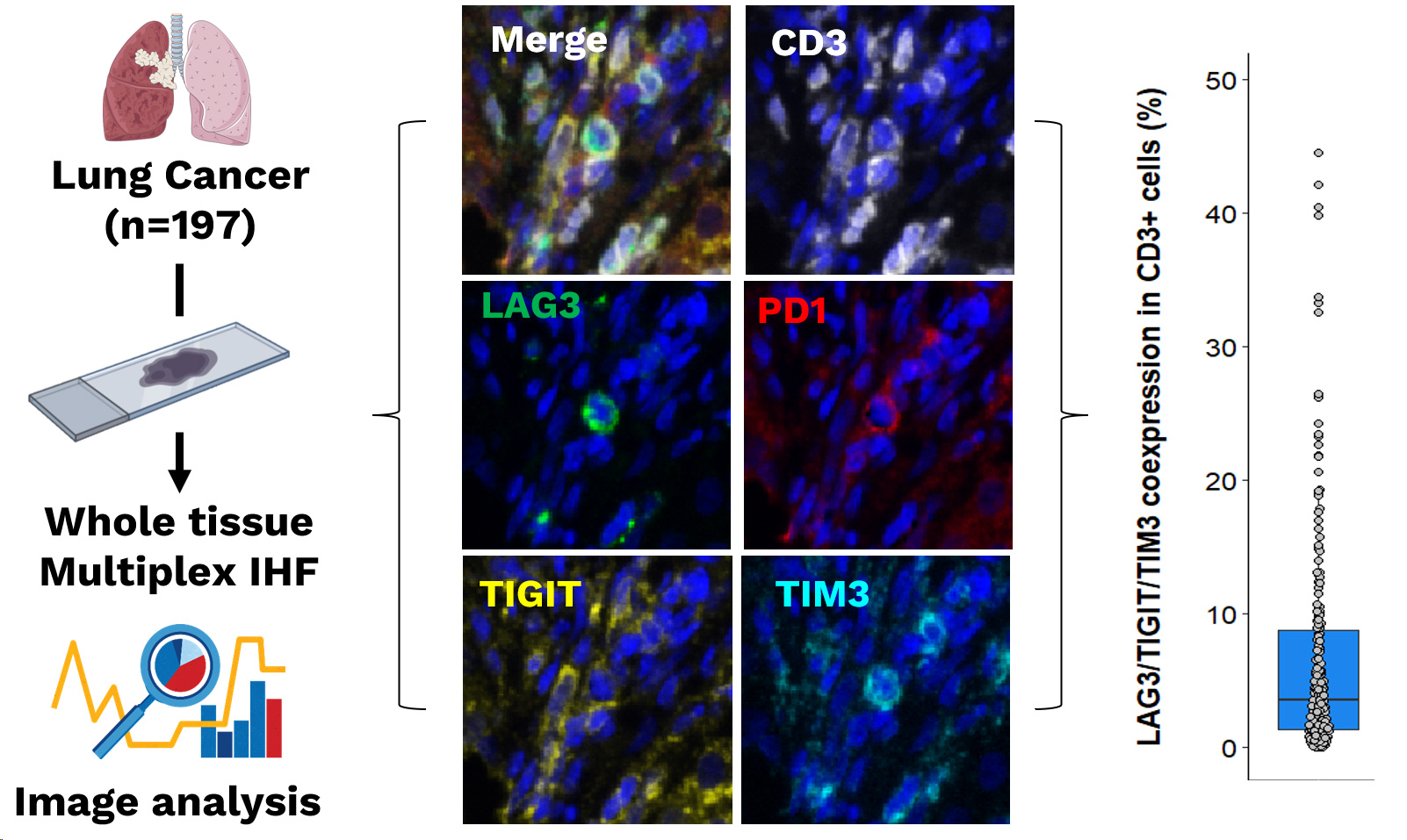
Detection of tumor-infiltrating CD3+LAG3+TIM3+TIGIT+ cells in Lung cancer patients. The presence of tumor-infiltrating lymphocytes expressing exhaustion markers was assessed on tumor samples from 197 Lung cancer patients using a multiplex immunofluorescence (mIHF) assay combining CD3, LAG3, PD1, TIGIT, and TIM3 markers. The images show the expression of each single marker assessed, with its corresponding fluorescence spectrum, and their combination (merge), respectively. Finally, image analysis allowed to define for each patient, the percentage of LAG3/TIGIT/TIM3 expressing cells within total CD3+ T cells, thus highlighting variability among patients.
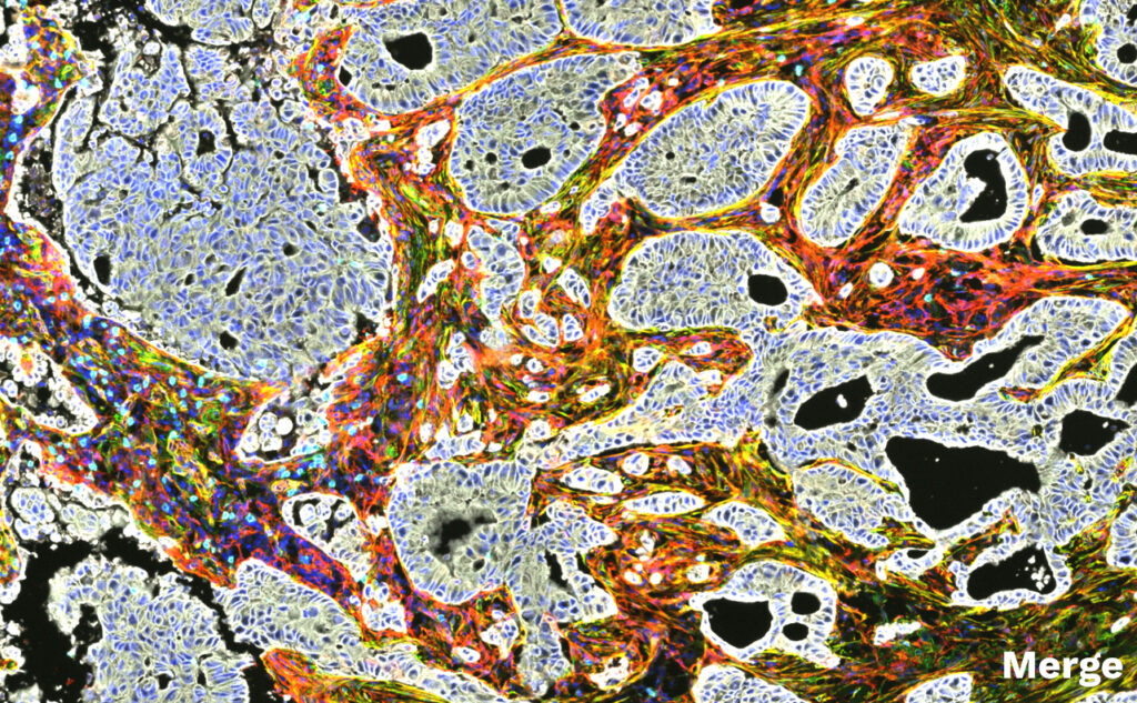
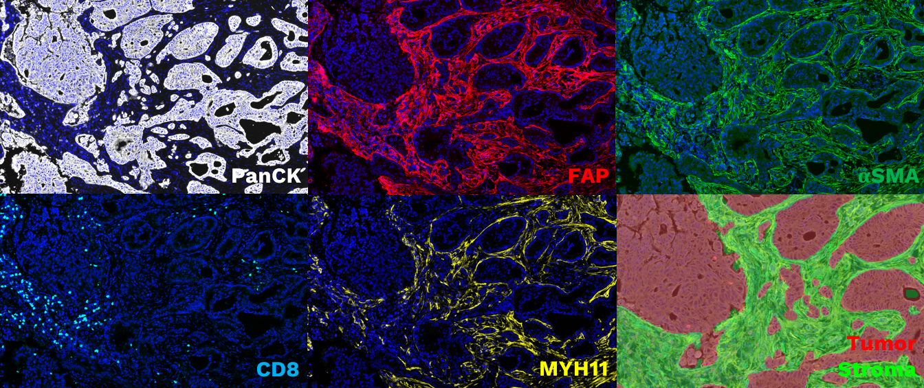
Detection of cancer-associated fibroblasts using a validated multiplexed immunohistofluorescence (mIHF) panel combining PanCK, CD8, FAP, MYH11, and aSMA markers. Representative image field of PanCK/CD8/FAP/MYH11/aSMA/DAPI multiplexed immunohistofluorescence panel on a FFPE NSCLC adenocarcinoma section. All scales, 5X. Illustration of the segmentation strategy of the tissue in “Stroma” and “Tumor” areas is shown at the bottom right.
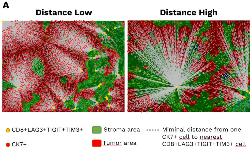
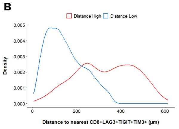
Spatial analysis of the CD8+LAG3+TIGIT+TIM3+ distribution in NSCLC patients. (A) Illustration of the calculated distances between epithelial tumor cells (CK7 positive) and tumor-infiltrating CD8+LAG3+TIGIT+TIM3+ cells. Tumor and Stroma areas are highlighted in red and green, respectively. (B) Density plot of the median distances of CK7+ to nearest CD8+LAG3+TIGIT+TIM3+ in patients classified as “Distance High” (red line) and “Distance Low” (blue line).
Your contacts

Talk to our team !
Paul Marteau, PharmD (preclinical study director), Imane Nafia, PhD (CSO), Loïc Cerf, MSc (COO), Alban Bessede, PhD (founder, CEO), Jean-Philippe Guégan, PhD (CTO)
Tell us about your project !
Digital pathology services I Immuno-Oncology CRO I Quantitative histopathology
Tissue-based biomarker investigation is a key step in pre-clinical / clinical drug development as it can serve either identification of novel therapeutic target, assessment of a surrogate marker of drug efficacy as well as prediction of a candidate compound benefit. To support novel therapies development at both pre-clinical and clinical level, we offer expert histology services relying on automated and standardized stainings capabilities spanning from monoplex to 7-plex marker panels and using either conventional chemistry or fluorescence revelation systems (immunohistochemistry/immunohistofluorescence). Many types of samples – either tumor or normal tissue, originating from human or pre-clinical models – can be adressed using our platform. Multispectral images acquired on our PhenoImager HT (Akoya Biosciences) are processed through image analysis to provide quantitative data including phenotyping, marker expression, as well as geographical distribution of cells within the samples. In addition, bespoke services can also be offered to specifically meet our clients’ needs. Key advantages of tissue-based imaging: Exploration at multiple scales, from cell-to-cell interactions to the macroscopic tissue architecture. Well-suited for simultaneous detection of multiple markers to identify specific cell subsets, document an immune response or quantify biomarkers of interest. Automated and robust workflow from sample processing to multiplexed image acquisition and quantification.

