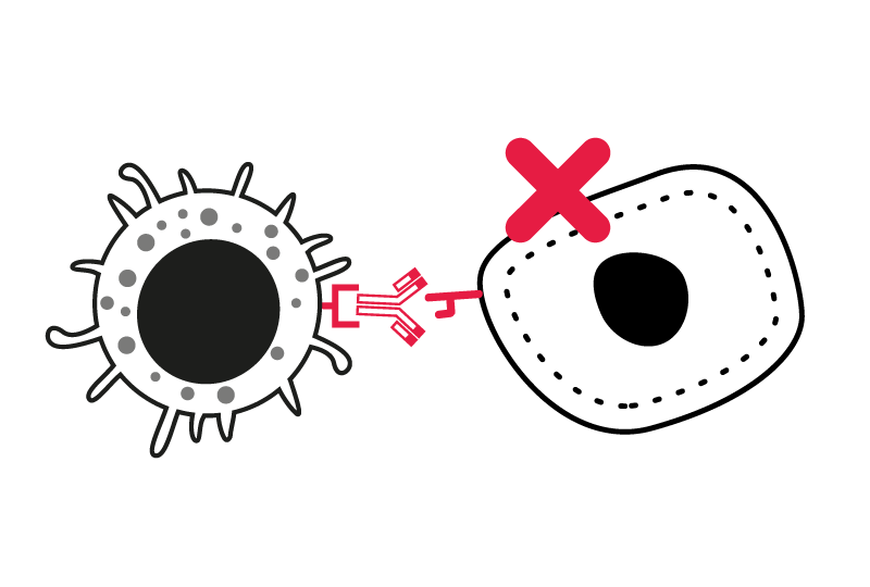
Immune cell-mediated tumor killing constitutes the most relevant functional assay to evaluate the effectiveness of candidates aimed at promoting the functions of effector cells against cancer cells.
Key features of our specialized killing assays:
- Human tumor cell lines, cultured with activated primary immune cells – PBMCs, or specific immune subsets such as T-/NK-cells directed or not against specific tumor antigens
- Tumor cell apoptosis monitored over time by live-cell imaging
- Immune cell response captured through the measurement of key cytokines
- Additional readouts available flow cytometry immunophenotyping, cytokine profiling, proteomics, transcriptomics
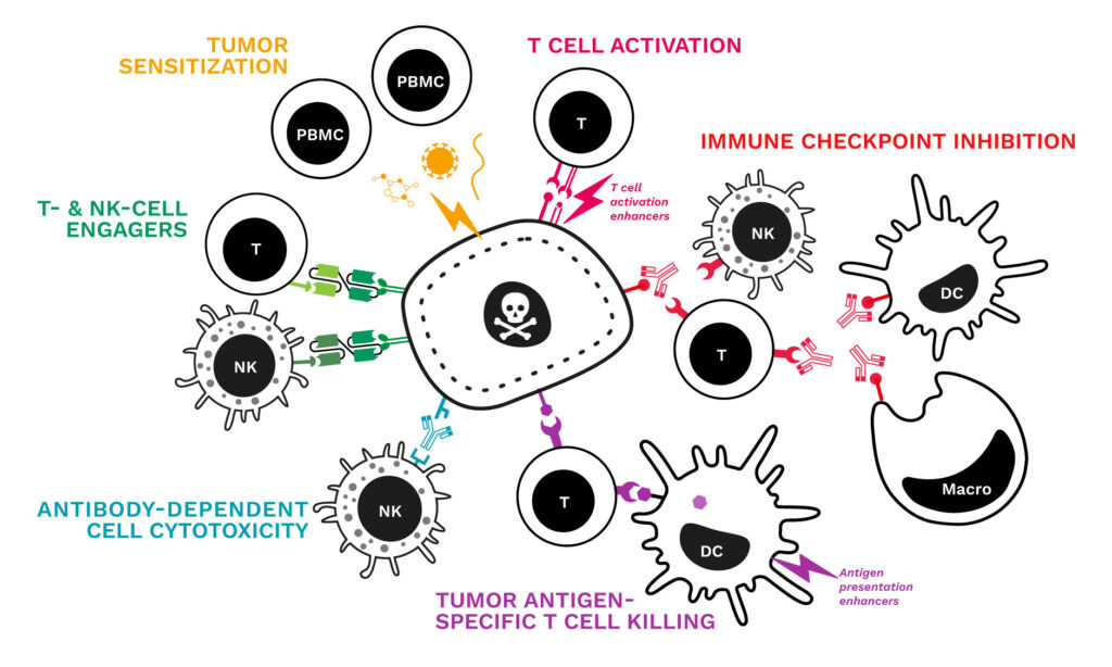
Our specialized tumor-killing assays enable the assessment immuno-oncology agents, including monoclonal antibodies, engagers, checkpoint blockers, antibody-drug conjugates, immunostimulatory agents, adjuvants (like TLR agonists), small molecule immunomodulators, bacterial toxins/superantigens, tumor-associated antigens and immunogenic peptides, and cytotoxic compounds either acting directly or by triggering immune cell activation (chemo, targeted therapy drugs).
Why working with Explicyte?
Experts
in Immuno-Oncology
- 100+ in vitro campaigns conducted over the past 10 years
- 30+ peer-reviewed publications in key immuno-oncology journals
- A comprehensive technology platform to decipher mechanisms of action (activation markers, signaling pathways…)
Personalized
approach
- Targeted discussion based on your request to design bespoke strategy & fit-for-purpose study proposal
- A dedicated study director (PhD level) from experimental plan to final report discussion
- Custom cellular models: over 100 human cancer cell lines available, to be cultured with target primary immune cells
Tell us about your project !

Talk to our team !
Paul Marteau, PharmD (preclinical study director), Imane Nafia, PhD (CSO), Loïc Cerf, MSc (COO), Alban Bessede, PhD (founder, CEO), Jean-Philippe Guégan, PhD (CTO)
Immune cell-mediated tumor killing assays for cancer immunotherapy I CRO services
Explicyte Immuno-Oncology provides functional cellular assays to assess the killing efficacy of T cells and Natural Killer (NK) cells. Target tumor cell killing is measured over time, using timelapse imaging (Incucyte) and fluorescent probes to monitor tumor cell number and apoptosis. Various key immune cell subsets play an important role in mediating tumor cell death through several ways involving immune-mediated cell killing mechanisms such as T cell cytotoxicity-mediated killing and antibody-dependent cell cytotoxicity (ADCC), which result in the induction of target cell apoptosis. One of the current promising challenge in cancer immunotherapy is the potentiation of the specific elimination of tumor cells. While in vitro immune cell killing is recognized as perhaps one of the most relevant functional measures to evaluate the ability of candidate compounds or antibodies in promoting the effector immune function, many techniques have been developed and appropriate in vitro assays for the evaluation of the effectiveness of immunotherapeutics must more and more fulfill requirements in terms of high sensitivity and quantitative analysis, and should also allow the analysis of the tumor cell death occurring within days instead of hours. Explicyte is offering specialized in vitro T cell cytotoxicity-mediated killing assay and ADCC assay to evaluate the immunomodulatory function of test compounds / antibodies. Our sensitive and quantitative assays based on the use of 96-well plate co-culture of human tumor cells* and primary immune cells along with a live cell imaging platform enables the kinetic monitoring of the killing immune response towards tumor cells and thus analysis of cell death specifically within the tumor cell population. Explicyte is offering a specialized in vitro T cell cytotoxicity-mediated killing assay to evaluate the ability of test compounds to modulate the immune response of effector cell subsets. Our sensitive and quantitative assay is based on a 96-w plate co-culture of human tumor cells* and primary immune cells and consists in the kinetic monitoring of tumor cell proliferation and death thereby enabling to measure the killing immune response towards tumor cells by analyzing cell death specifically within the tumor cell population. Understanding the processes of the immune system and its interactions with tumor cells and gaining deeper insights into mechanisms involved in their destruction are essential to identify and validate new targets and assess novel therapeutic approaches. While one of the promising cancer immunotherapy challenges is the potentiation of immune cell attacks of tumor cells, in vitro immune cell-mediated killing is recognized among the most relevant functional measures to evaluate the ability of lead items to promote the functions of effector immune cells, thereby representing a valuable tool for immuno-oncology drug discovery.
We designed specialized in vitro immune cell-mediated killing assays to assess immunomodulatory lead drugs for their ability to enhance the immune response against tumor cells. While based on co-cultures of human immune cells and target tumor cells, our assays allow for the parallel capture of the immune response and tumor sensitivity. Interestingly, they can thus be used to evaluate lead drugs for their potential to enable tumor cell destruction by immune cells according to various possible immunotherapy strategies: by blocking immune checkpoints; by activating T cells (via TCR ligation or involving other (co)stimulatory receptors); by bridging immune cells to their targets thus physically engaging immune and tumor cells to redirect the response;through ADCC mechanisms; by enhancing DC-mediated tumor antigen presentation for the priming of tumor antigen-specific cytotoxic lymphocytes and induction of an adaptive effector T cell-mediated killing response.


