Recent studies have nicely demonstrated the participation of B cells in the response to immunotherapies in different cancer indications including Melanoma1, Renal Cell Carcinoma2 as well as Sarcomas3. Indeed, a strong B cell signature in samples from responding patients, when compared to the “non-responders”, has been highlighted – B cells being particularly enriched within organized lymphoid aggregates known as Tertiary Lymphoid Structures (TLS). Mature TLS harboring a germinal center favor B cell development and function and thus participate to the initiation and regulation of an anti-tumor immune response. Finally, the fact that TLS presence before treatment initiation is predictive of a better clinical outcome brings the B cells to the forefront of cancer immunotherapy4 and warrants much more investigations.

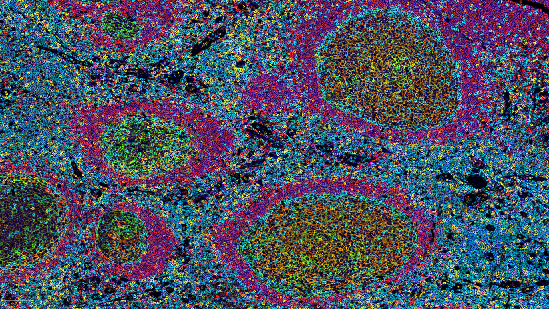
As to participate and support B cell research in cancer immunotherapy, we set up an histological TLS analysis based on i) automated stainings using multiparametric immunohistofluorescence analysis combining CD4, CD8, CD20, CD21, and CD23 markers, and on ii) staining digitization and image analysis as to get quantitative data (TLS size & density, mature vs immature…). Customized panel(s) and image analysis can also be applied as to better characterize TLS biology in the context of specific projects.

Learn more about our capacities for TLS staining & scoring
1 Jonsson et al., Nature. 2020 Jan; 577(7791):561-565
2 Wargo et al., Nature. 2020 Jan; 577(7791):549-555
3 Friedman et al., Nature. 2020 Jan; 577(7791):556-560
4 Tulia C. Bruno, Nature 2020, Jan; 577, 474-476