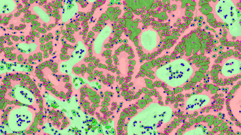
To deal with the current challenge of a qualitative and quantitative tumor immune landscape characterization, we have set up a fully integrated platform spanning from FFPE slide preparation and staining, to image digitization and analysis.
As prototypical immunotherapies, immune checkpoint inhibitors (ICI) targeting PD-1 / PD-L1 axis and CTLA-4 have achieved unprecedented clinical efficacy thereby shedding light on the crucial role of the tumor microenvironment and immune landscape in the control of tumor progression. Even not yet fully understood, benefit of ICI depends on several parameters including intratumoral immune contexture.
To deal with the current challenge of a qualitative and quantitative tumor immune landscape characterization, we have set up a fully integrated platform spanning from FFPE slide preparation and staining, to image digitization and analysis. This combined approach – taking advantage of a series of validated markers – can be applied in monoplex or multiplex mode through conventional immunohistochemistry (IHC) or immunohistofluorescence (IHF). Our platform thus provides the ability to delineate novel predictive biomarkers as well as insights into mechanisms of action of innovative cancer immunotherapies based on the characterization of the human immune contexture (cell density, immune cell proximity to tumor cell, etc.).
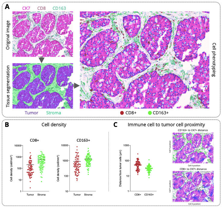
Multiplexed immunohistochemistry imaging for immune contexture analysis in human lung adenocarcinomas.
(A) A human FFPE section was automatically stained on a Ventana discovery platform using a combination of antibodies targeting Cytokeratin 7 (CK7, for tumor cells, pink), CD163 (for tumor-associated macrophages, green), and CD8 (for CD8 T cells, brown). Image was originally acquired through a multispectral imaging system (Vectra® Polaris™, Akoya Biosciences), and then processed through inForm® analysis software (Akoya Biosciences) and machine learning approach for tissue segmentation (tumor and stroma regions) and then for cell segmentation & phenotyping.
(B) Immune cell density (CD8+ and CD163+) quantification for each patient (n=80) within the different tissue regions highlighted a highest immune cell density within the stroma compared to the tumor. In addition, this analysis revealed an expected heterogeneous infiltration profile that could underlie sensitivity to therapies.
(C) In addition to cell density analysis, proximity of immune cells (CD8+ or CD163+) from tumor cells (CK7 positive) has been calculated for each patient (n=80) and, again, demonstrated a patient-dependent pattern.


