
Large-cohort spatial studies have long forced a tradeoff between transcriptome breadth, single-cell resolution, and throughput. Atera, 10x Genomics' new in situ spatial platform, is built to remove that constraint— and Explicyte is opening a limited number of early Atera slots for 2026 (1 to 4 slides per slot), and you can reserve yours now.
Atera measures the whole transcriptome directly in intact tissue, at single-cell resolution, across both human FFPE and fresh-frozen samples. For discovery and translational programs, that means:

You may be a good fit for an early Atera slot if you want to test whole-transcriptome depth early on:
Reserving now secures capacity in the 2026 queue and lets us scope your project ahead of time.
What Atera brings
Atera measures the whole transcriptome directly in intact tissue, at single-cell resolution, across both human FFPE and fresh-frozen samples. For discovery and translational programs, that means:
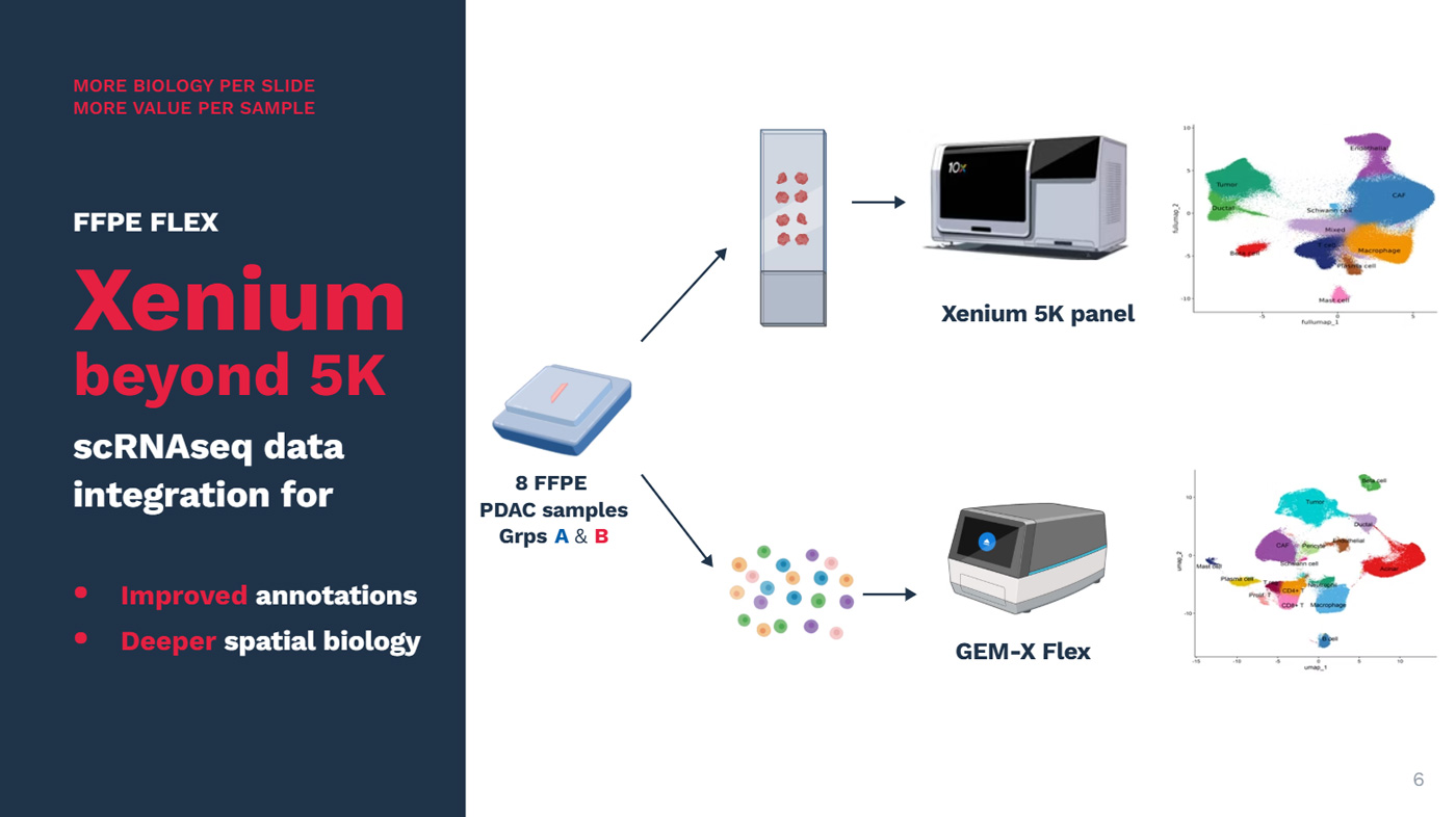
- Whole-transcriptome depth — profile the full transcriptome spatially (>18K genes), rather than a fixed target panel.
- Single-cell resolution — resolve expression cell by cell within the tissue architecture.
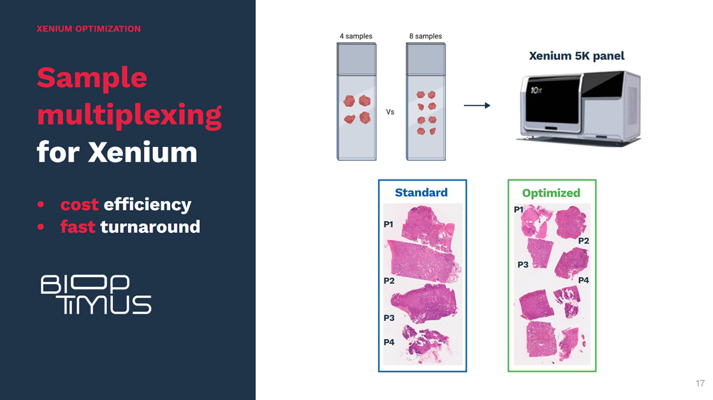
- Cohort-scale throughput — 4 slides per run, to move from single specimens to full cohorts.
"Atera is a step change in spatial transcriptomics we weren't going to wait on. It pairs whole-transcriptome depth and single-cell resolution with the throughput to run large cohorts — which is exactly what clinical research and AI-ready dataset generation demand. For that work, I see it becoming the reference technology."
— Alban Bessède, PhD, CEO, Explicyte
Why Explicyte for your first runs on Atera
- Trusted 10x Genomics service provider Explicyte is a certified 10x Genomics service provider across Xenium, Visium HD, and Chromium X, with the workflow experience to bring the same standard to Atera.
- Easy logistics for EU samples Based in France, we make sample shipping and return straightforward for European teams. We can also help source cohorts of interest through our French biobank network.
- Expert sample prep and QC Our pathologists handle histopathological review, and we run systematic RNA quality control so nothing is wasted on unusable material.
- Flexible pilot slots While standard Atera runs typically comprise four slides, Explicyte gives you the option to run a single slide in 2026, enabling you to evaluate the technology cost-effectively before scaling up.
- In-house data science team Our data science team built a proprietary workflow for QC, cleaning, annotation, and analysis of 10x Genomics datasets — ready to apply to your Atera data.

Is this for you?
You may be a good fit for an early Atera slot if you want to test whole-transcriptome depth early on:
- Human FFPE tissue or tissue microarrays
- Human fresh-frozen tissue
Reserving now secures capacity in the 2026 queue and lets us scope your project ahead of time.


 Promotes Tumor Progression and Metastases in Undifferentiated Pleomorphic Sarcoma")


