

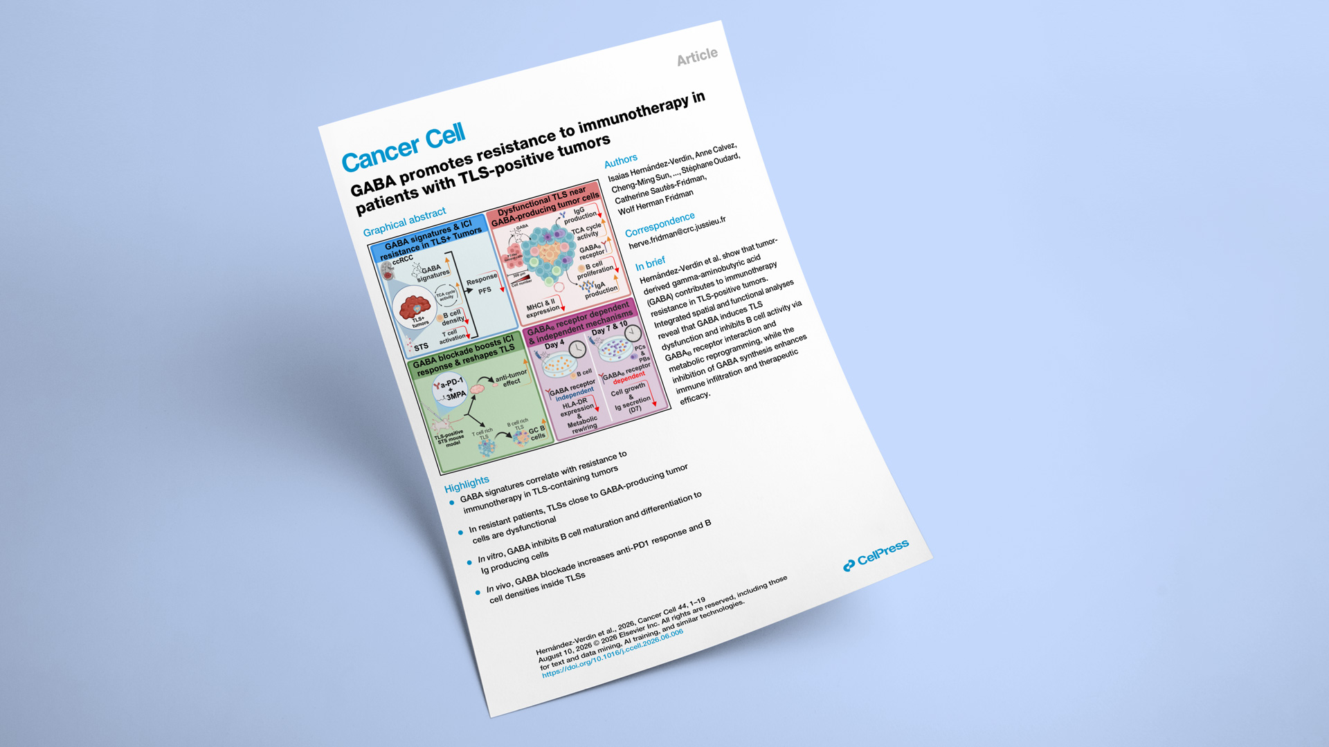
Explicyte contributed to a newly released Cancer Cell paper, which identifies tumor-derived GABA as a driver of immunotherapy resistance in TLS-positive tumors. By integrating renal cancer and soft-tissue sarcoma cohorts with spatial transcriptomics, metabolomics, tissue imaging, human B-cell assays, and an immunocompetent sarcoma model, the team showed that GABA disrupts B-cell activity and TLS function. Blocking GABA synthesis enhanced anti-PD-1 efficacy and increased B-cell proliferation within TLSs, supporting a new biomarker and therapeutic strategy for ICI-resistant tumors.
Explicyte co-authors Alban Bessede and Jean-Philippe Guégan contributed to transcriptomic analysis in this Cancer Cell paper. The study also incorporated the TLS-selected PEMBROSARC soft-tissue sarcoma cohort and used the Immusmol anti-human GABA antibody IS1006 for tissue analyses. Led by the Centre de Recherche des Cordeliers, the collaboration connected renal cancer and sarcoma clinical cohorts with spatial and functional models of B-cell immunity.
The question: Why do some TLS-positive tumors resist immune checkpoint blockade — and can targeting GABA restore response?
KEY STEPS
Define a GABA resistance signature
The team analyzed 58 TLS-positive tumors from ICI-treated patients in the BIONIKK clear cell renal cell carcinoma cohort, including 31 responders and 27 nonresponders. Screening 9,321 MSigDB signatures plus 33 curated signatures identified GABA synthesis, transport, secretion, and receptor pathways among the strongest features of resistance. Using the GABA ccRCC signature, 85% of GABA-high patients were nonresponders versus 26% of the GABA-low group (P = 2.39 × 10⁻⁶), with significantly shorter progression-free survival (P = 0.0069).
Map dysfunctional TLS neighborhoods
Visium spatial transcriptomics profiled 19 BIONIKK tumors — 10 from nonresponders and nine from responders — alongside multiplex staining in 58 tumors and highplex immunofluorescence in nine. GABA synthesis localized mainly to ACE2-positive, proximal tubule-like malignant cells. Across 109 TLS-annotated regions, TLSs from nonresponders showed reduced plasma-cell, IgG, antigen-presentation, and cytotoxic programs, while GABA-receptor and TCA-cycle activity increased.
Test GABA on human B cells
Purified human B cells were differentiated toward plasmablasts and plasma cells in the presence of 100–1,000 μM GABA. Across five donors and six experiments, GABA reduced HLA-DR expression at day 4 (P = 0.016), while later assays showed lower proliferation and reduced IgM and IgG secretion without a corresponding loss of viability. The GABAB-receptor antagonist saclofen partially rescued cell growth and immunoglobulin production, supporting both receptor-dependent and metabolic mechanisms.
Validate the signal in sarcoma
The PEMBROSARC Strate 6 analysis included 13 patients with metastatic, TLS-positive soft-tissue sarcoma treated with pembrolizumab and low-dose cyclophosphamide. A 13-gene GABA STS signature containing GAD1 and GABA-receptor genes was significantly enriched in nonresponders (P = 0.0016). All seven GABA-high patients were nonresponders, compared with one of six GABA-low patients (P = 0.0046), with a trend toward shorter progression-free survival (P = 0.077).
Block GABA synthesis in vivo
In the TLS-forming MCA-OVA syngeneic sarcoma model, the GAD67 inhibitor 3-mercaptopropionic acid had little effect alone but enhanced anti-PD-1 activity. The combination reduced tumor weight by approximately 75% versus control, and B-cell depletion weakened this benefit. Compared with anti-PD-1 alone, combination treatment increased the intra-TLS B-to-T-cell ratio from 2.68 to 8.07 and raised proliferating Ki67-positive B-cell density 3.7-fold (P = 0.019).
Impact
GABA emerges as more than a correlative biomarker: it is a spatially organized metabolic resistance mechanism that suppresses B-cell function inside tumor-associated TLSs. The findings also show why detecting TLSs alone may not be sufficient to predict immunotherapy response.