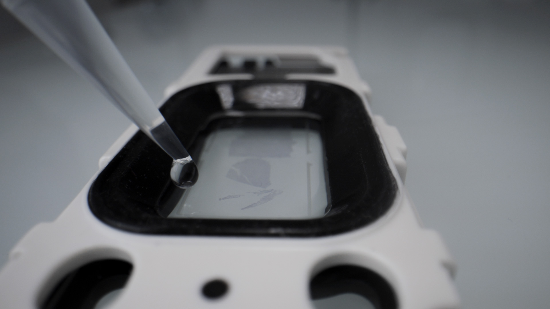
Formalin-fixed, paraffin-embedded (FFPE) tissues remain the backbone of oncology biobanking, with vast archives of clinically annotated samples. With 10x Genomics’ probe-based workflows —Chromium Fixed RNA Profiling, Visium FFPE, and Xenium—these archives are now tractable for robust transcriptomic profiling to uncover disease mechanisms and identify new drug targets and biomarkers.
One of the first CRO in Europe equipped with the full 10x suite (Chromium X, Visium HD, Xenium), Explicyte is a 10x-certified provider specializing in precision oncology. For the past decade, our team has leveraged FFPE specimens for translational studies end-to-end—from custom tumor tissue sourcing and QC, to data generation, analysis, and presentation in sponsor reports and peer-reviewed publications.
Below, we share our key insights for FFPE transcriptomics—how to qualify samples, choose the right platform according to the question, and integrate multi-scale data to drive translational decisions.
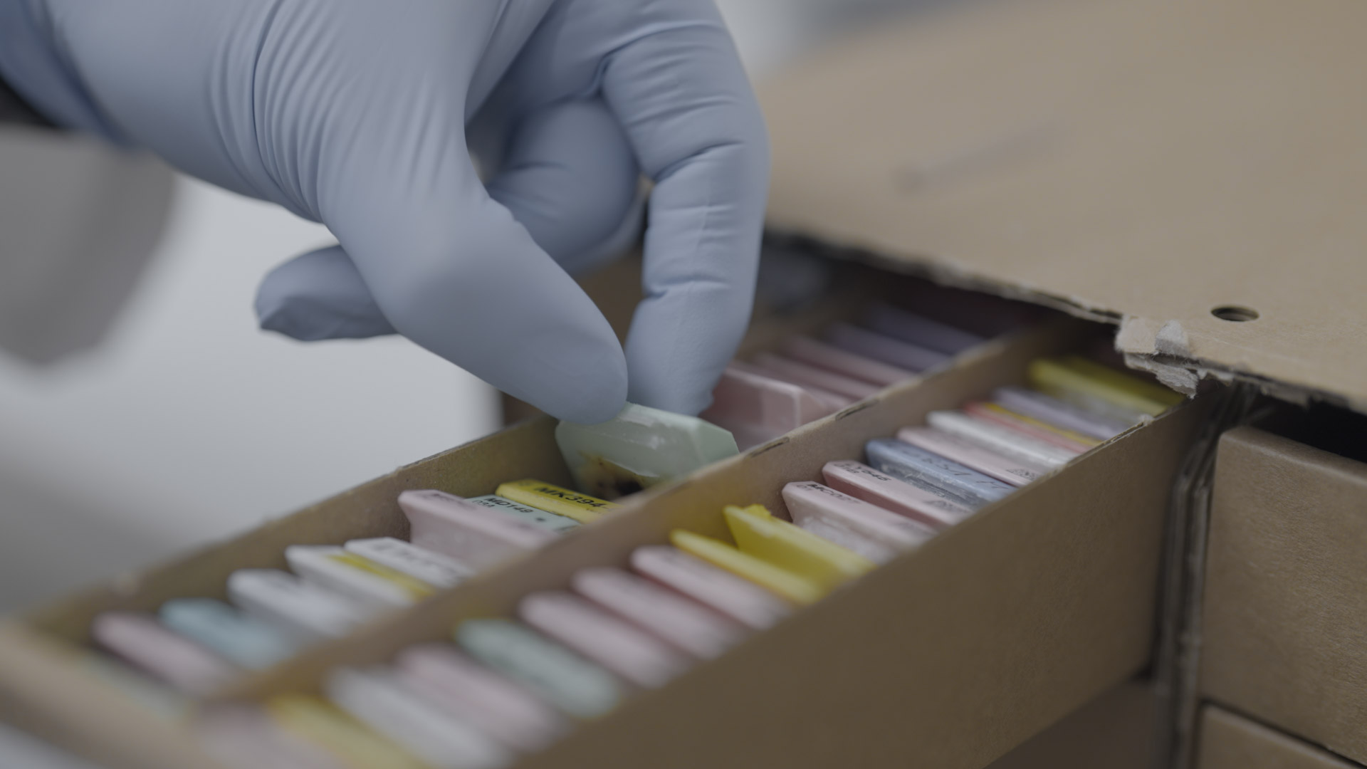
Sample qualification comes first
Transcriptomic studies are a significant investment—so every sample must count. The design of the tissue collection and the inclusion/exclusion criteria determine both the scientific value and the cost-efficiency of the final dataset.
Sourcing
- We partner with accredited French biobanks to source FFPE specimens that match the target indications, prior treatments, and required clinical metadata (e.g. treatment exposure, PD-L1 score, survival endpoints).
- Each specimen enters our QMS for tracking.
Pre-analytical QC
- RNA integrity: RNAscope staining (privileged) or DV200 assessment.
- Pathology review: we work with consulting pathologists to confirm tumor content (≥20–30%), necrosis, inflammation/TLS status, and viable tissue area adequate for the planned workflow.
- Block history: we exclude over-fixed, old/degraded, heavily pigmented or decalcified samples.
- Sectioning & storage: we cut to optimal thickness (typically 5 µm), use freshly cut sections when possible, and store under controlled conditions to minimize oxidation/hydrolysis before runs.
Once the FFPE inputs pass QC, we can proceed to platform selection with confidence.
Platform selection is about the question (and time/budget constraints)
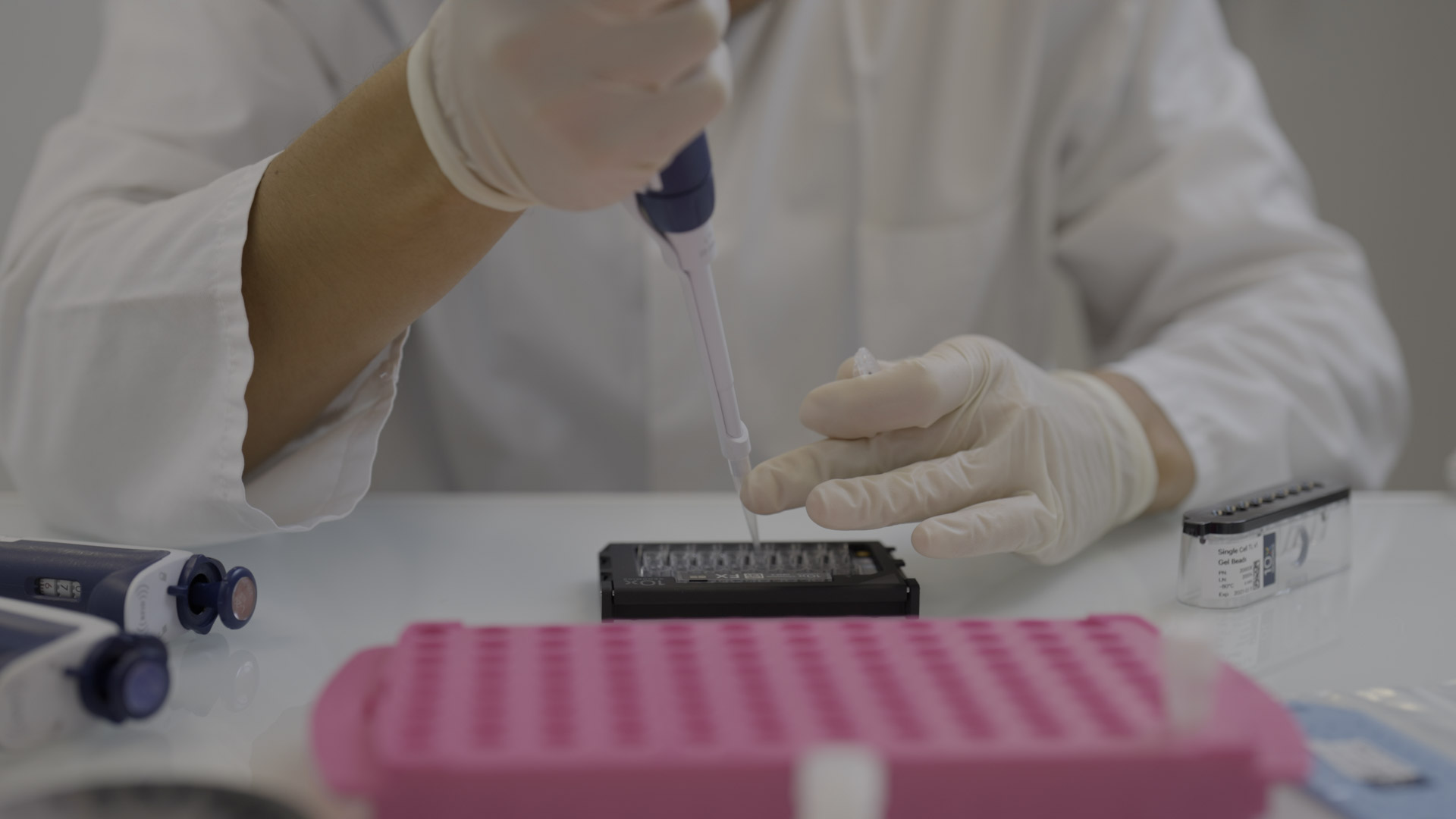
Chromium X — fast, multiplexed single-cell RNA-seq (no spatial information)
What it delivers:
Single-cell (or single-nucleus) transcriptomes from human FFPE using a probe-based whole-transcriptome panel (~18k genes).
Why use it:
- Scalable cell-type/state discovery across cohorts
- Comparative studies between groups (e.g., pre- vs. on-treatment, responders vs. non-responders) and pathway enrichment
- High-throughput multiplexing with sample tagging—up to 3,072 samples per chip/run—reducing cost per sample and time-to-data
Trade-offs:
- Destructive and no spatial localization: you learn who is present and what they express, not where they reside in tissue
- Rare populations: practical input/recovery per sample (tens of thousands of cells/nuclei) can limit detection of rare cell types; consider enrichment or deeper sequencing
- Sequencing step required: turnaround and cost depend on sequencing depth and queue
Great for:
Preliminary cohort screening, target/biomarker nomination, and deep cell-state biology when spatial context is secondary or slated for later in-situ validation.
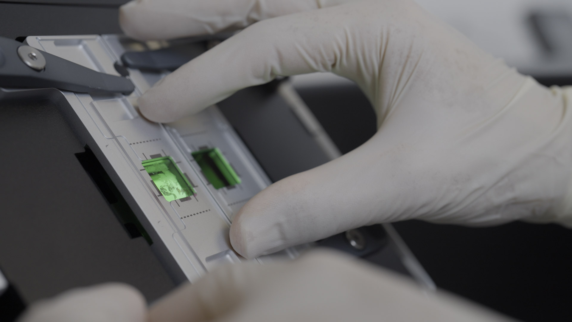
Visium HD for FFPE — whole-transcriptome, high-density spatial maps
What it delivers:
Whole-slide, spatially resolved transcriptomics on FFPE with a dense feature grid, co-registered to H&E (and optional IF) for histology-anchored analysis.
Why use it:
- Whole-tissue coverage to map regional programs (tumor core vs. margin, stroma, TLS, necrosis, etc.) with transcriptome-wide breadth
- Practical throughput (e.g., two tissue sections per slide) for hypothesis generation and tissue-scale patterning
Trade-offs & caveats:
- Higher resolution than legacy spot-based Visium, but not truly single-cell everywhere; partial cell mixing can occur in dense regions
- Adipose-rich or fragile tissues may shift during processing, causing minor mis-registrations between histology and expression layers
- Not all indications are equally tractable (e.g., decalcified bone; heavily pigmented/melanin-rich areas require caution)
Great for:
Tissue-level atlases, discovery of spatially organized programs, and selecting ROIs/genes for subsequent in-situ validation.
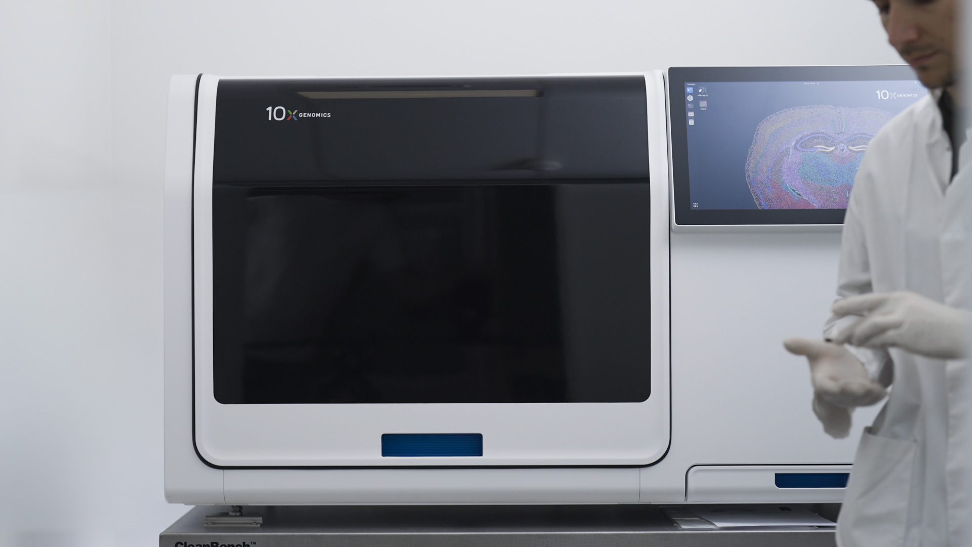
Xenium for FFPE : Single-cell & subcellular spatial transcriptomics with targeted panels
What it delivers:
Imaging-based in situ transcriptomics at single-cell (often subcellular) resolution on FFPE. Predefined panels (hundreds to ~5,000 genes) + custom add-ons (up to 100); protein co-detection available.
Why use it:
- True cellular neighborhoods: Quantify cell–cell interactions across all lineages (Tregs, CD8⁺ T cells, dendritic cells, cancer-associated fibroblasts, myeloid cells) within the tumor core, at the invasive margin, and in structured niches such as TLS.
- Precise target validation: confirm expression patterns from Chromium/Visium with exact localization.
- Flexible study design: cost-efficient ~300–500-plex panels for focused questions; 5K panels for broader discovery-plus-validation; custom add-ons for program-specific genes.
- Throughput options: multiple tissue sections can be placed on a slide and/or multiplexed with sample tags to optimize per-run economics (exact layout depends on tissue size and panel).
Trade-off:
Targeted (not WTA), so panel design matters; we typically prototype with focused panels and scale to 5k or protein when needed.
Great for:
Spatial validation of drug targets/biomarkers, micro-architecture analysis (immune exclusion, TLS, tertiary structures), and mechanism-of-action studies.

Quick chooser (FFPE)
- Need breadth across many samples? → Chromium (single-cell, scalable multiplexing).
- Need whole-slide context at transcriptome scale? → Visium HD (spatial WTA, high density).
- Need cellular precision and interactions in situ? → Xenium (targeted panels, single-cell/subcellular).
A linear Chromium → Visium HD → Xenium sequence may look rational, but it’s rarely the most time- or budget-efficient. We favor a question-driven design: start from the sponsor’s hypotheses and the literature, choose the minimal mix of platform, panel, and multiplexing that delivers decisive evidence, and iterate only when the added value is clear.
Data analysis: one pipeline, three modalities
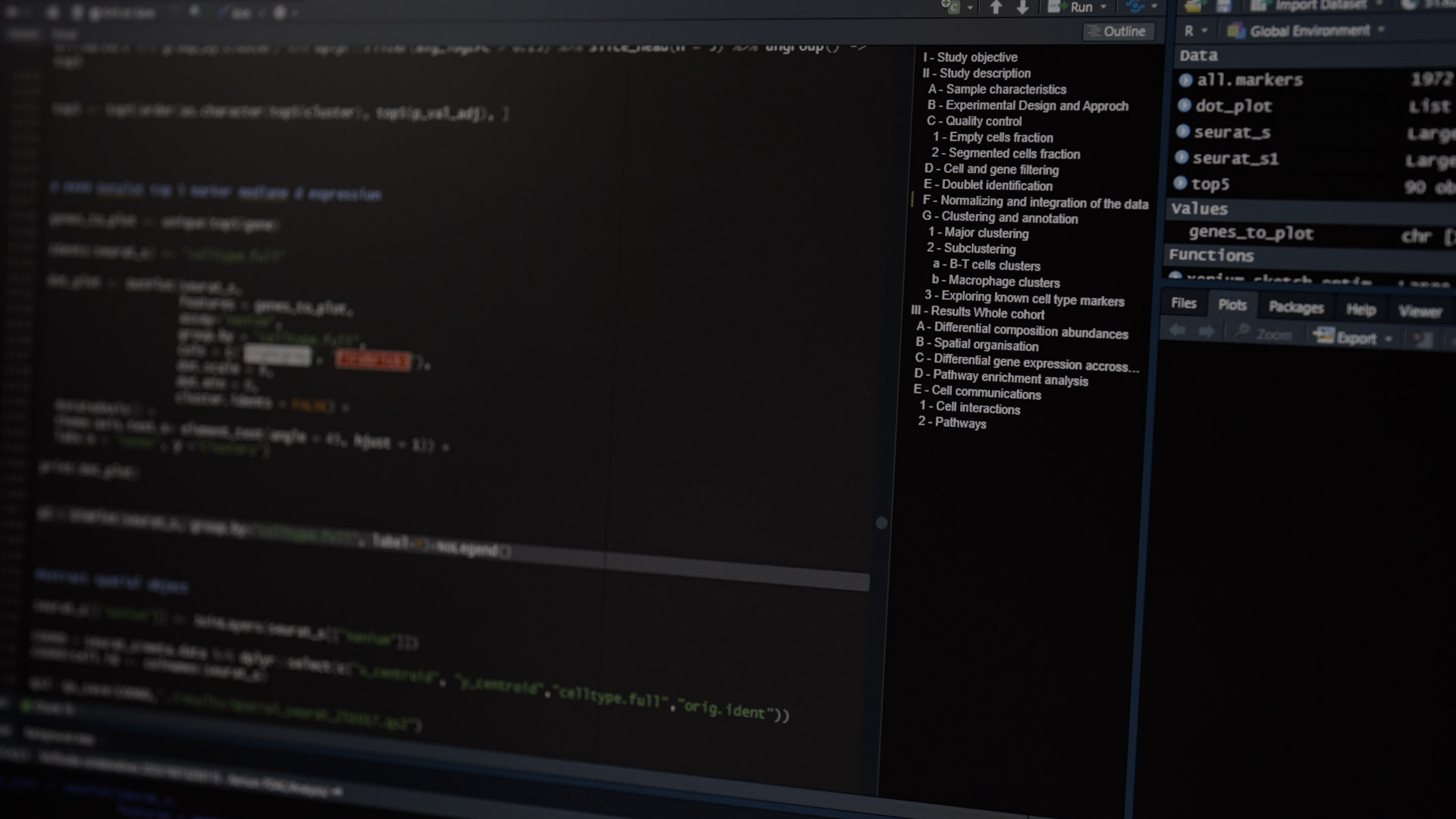
To keep cohorts consistent and comparable, we apply a unified analysis framework across platforms.
Core workflow
- Doublet mitigation, batch correction/integration
- Cell-type/spot annotation (reference-guided + de novo) and cell-type prioritization
- Pathway scoring and differential abundance/expression across clinical strata
Spatial analyses (Visium HD & Xenium)
- Neighborhood statistics and niche enrichment
- Targeted ligand–receptor inference
- Alignment to histopathology and IHF/IF imagery
Cross-platform integration
- We build cell-type references from Chromium and use them to deconvolve Visium and interpret Xenium
- Selection of Xenium gene panels from Visium/Chromium discoveries
- Multi-modal association with clinical covariates (e.g., PD-L1 TPS, TLS status, response, PFS/OS) with appropriate multiple-testing control
Deliverables
- Raw data and cleaned matrices, annotated objects (on request)
- Publication-ready figures/tables
- Concise reports mapping findings back to the original clinical questions
Learnings
Our team scaled from 2 PhD-level data scientists to 5 within a year. AI helps, but a biology-trained, critical eye remains essential to answer the right questions—and to discuss insights that surfaced in datasets.
Practical tips we’ve learned on FFPE tumors
- Panel strategy matters: Start targeted (cost-savvy) when hypotheses are clear; escalate to 5k Xenium or broader WTA when discovery is needed.
- QC saves budgets: A RNAscope gate and pathology-led triage prevent failed runs and misleading negatives.
- Mind the cohort design: Balance covariates to avoid confounding; record pre-analytical variables.
- Close the loop: Always plan in situ validation (IHF/IHC or Xenium) for high-value targets emerging from Chromium/Visium.
FAQ
Which platform should I use for FFPE when spatial context is critical?
Visium HD for transcriptome-wide, whole-slide context; Xenium when single-cell localization and cell–cell interactions are required.
What QC do you require for FFPE blocks?
RNAscope (preferred) or DV200 for RNA integrity; pathology review for ≥20–30% tumor content; checks for fixation age, pigmentation, and decalcification.
When should I choose a 5k Xenium panel vs a 300–500 gene panel?
Use focused panels for hypothesis-driven questions; escalate to 5k when broader discovery and validation are needed.
Do you support end-to-end studies from biobanking to reporting?
Yes—specimen sourcing and QC, platform runs, analysis, and sponsor-ready reports; raw/cleaned data and annotated objects available on request.

