Tissue segmentation
Machine-learning-driven analysis of tissue architecture based on pixel classification.
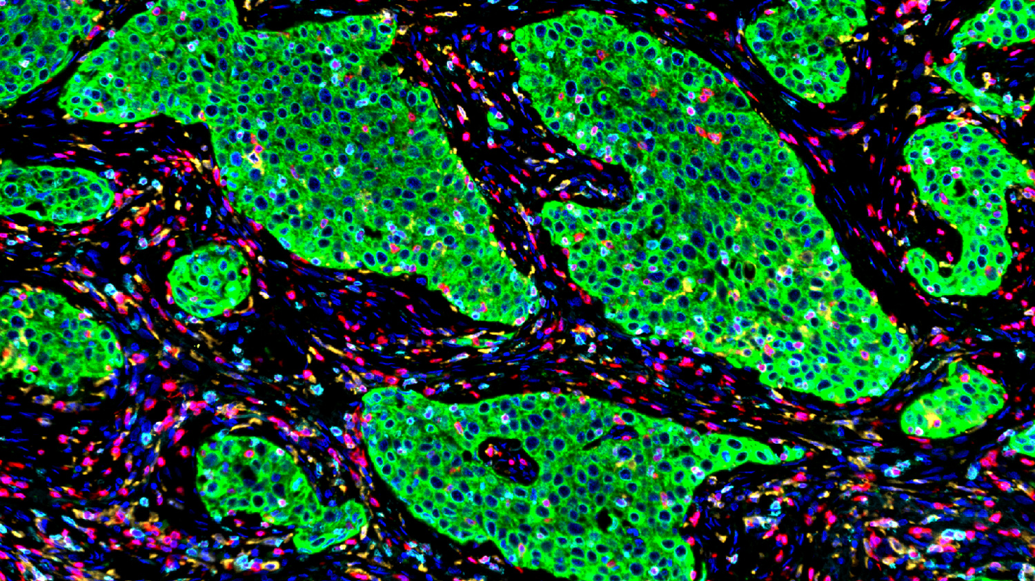
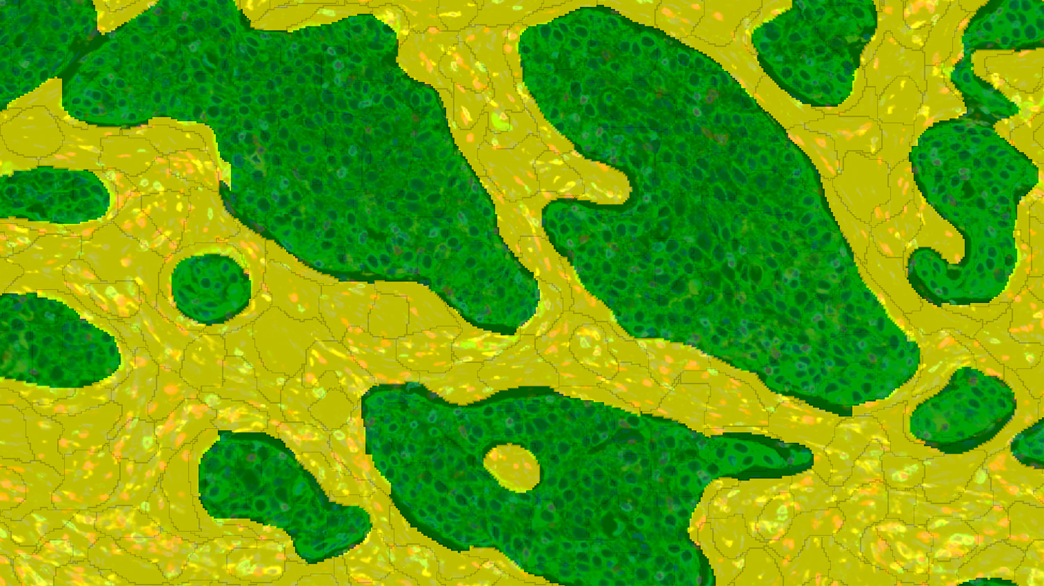
An NSCLC adenocarcinoma sample was stained using a multiplex immunofluorescence (IHF) panel: CD8 (cyan), CD4 (red), CD163 (yellow), PanCK (green), and DAPI (blue). The tissue was segmented into superpixels, classified automatically into Tumor (green) or Stroma (yellow) compartments using advanced machine-learning methods.
Cell segmentation & Phenotyping
Quantitative cell identification and characterization through proprietary segmentation workflows
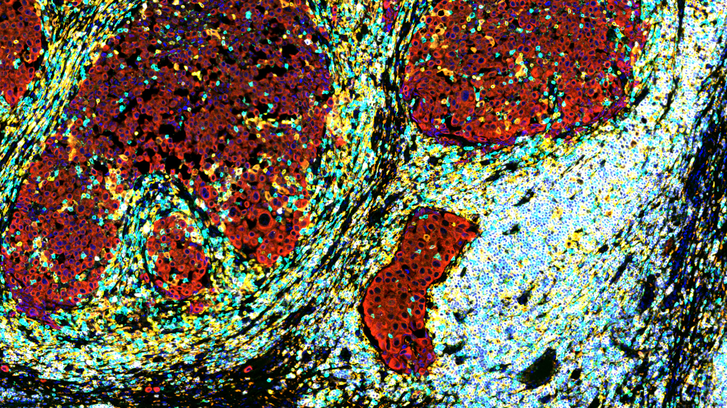
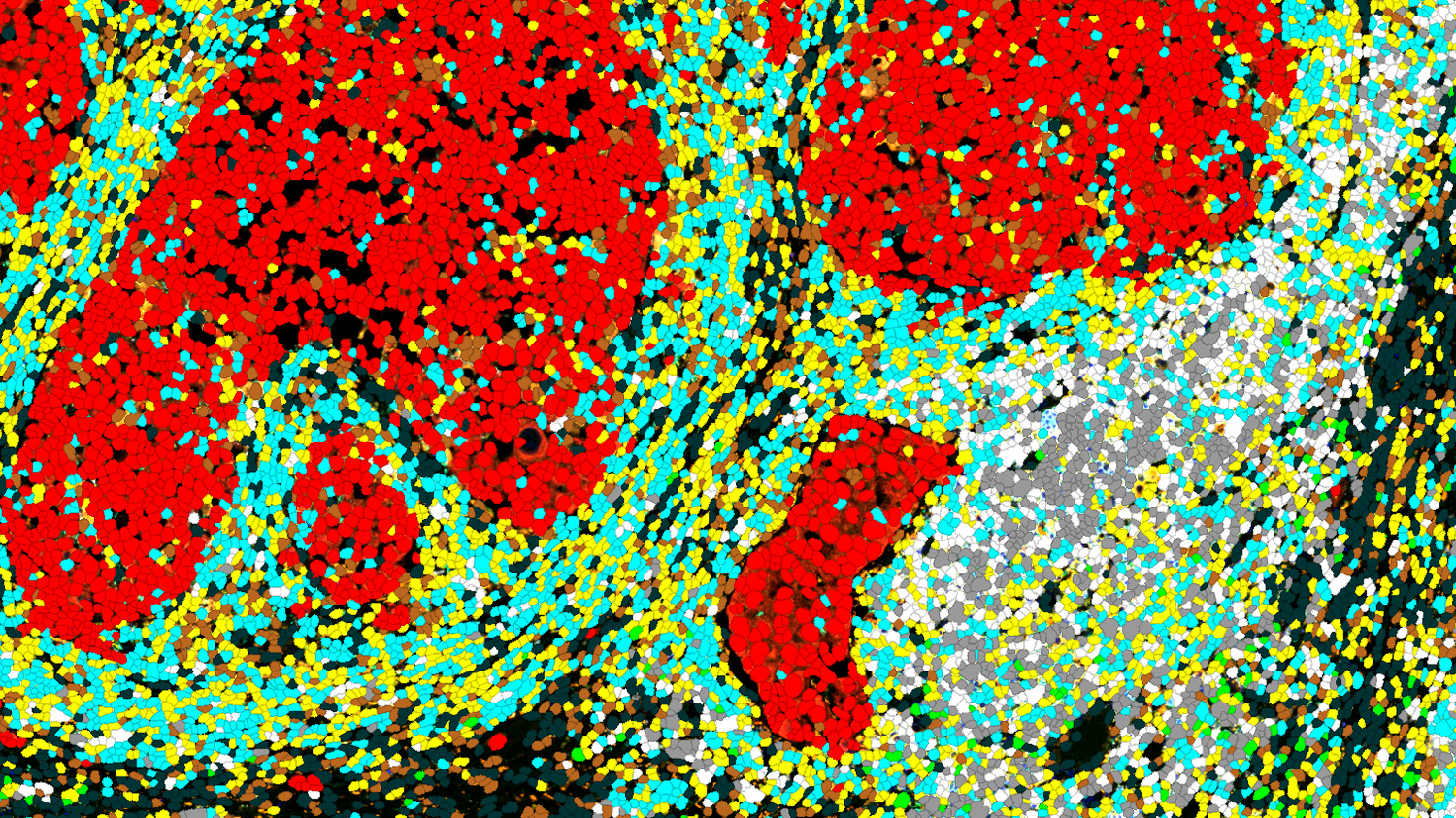
An NSCLC adenocarcinoma sample stained with a multiplex panel (CD3-yellow, CD8-cyan, CD20-white, CD23-green, CD163-orange, PanCK-red, and DAPI-blue) underwent individual cell segmentation. Following signal normalization, cell phenotypes were defined using a cytometry-based analysis workflow. Phenotypes included macrophages (orange), B cells (white), mature B cells (gray), follicular dendritic cells (green), CD4 T cells (yellow), CD8 T cells (cyan), and tumor cells (red). Unstained cells are shown in black.
Spatial analysis
Detailed analyses of spatial relationships, including nearest-neighbor distances, spatial heterogeneity, and cellular colocalization.
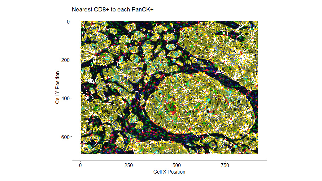
Nearest-neighbor distances were calculated between PanCK-positive and CD8-positive cells. The minimum distance between these cells is visually represented by white dashed lines.
Tailored bioinformatic analysis
In-depth computational analysis customized to your research goals, including marker expression profiling, object measurements, density quantification, and correlation with clinical metadata.
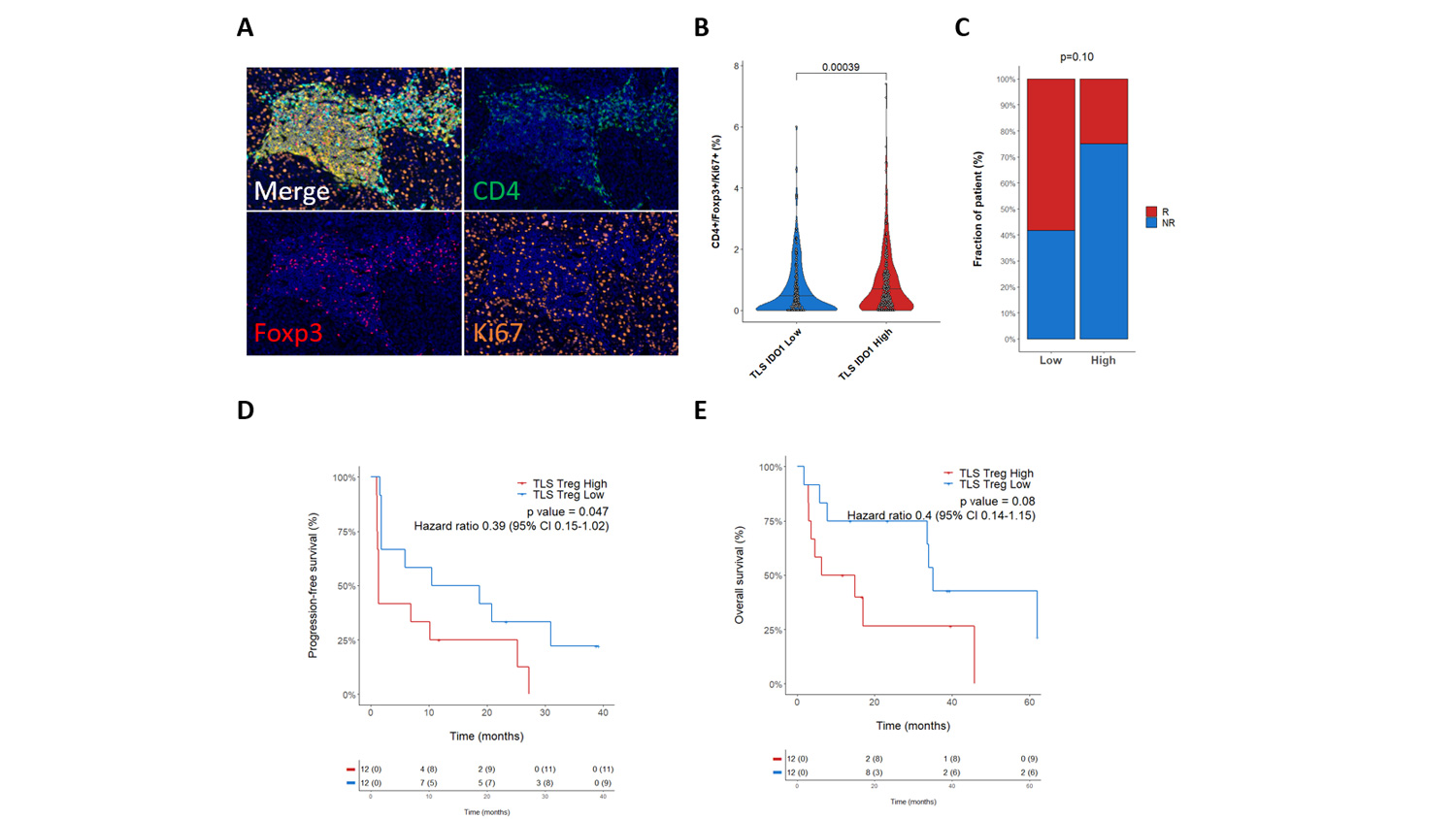
Bessede et al. Clin Cancer Res . 2023 Sep 26;29(23):4883–4893. doi: 10.1158/1078-0432.CCR-23-1928

Scientific interaction
A PhD-level data scientist in charge of your study discusses with your team your needs and expected deliverables.

Flexible data transfer
We provide a secure server for uploading your digitized slides and clinical metadata, and we can facilitate the physical transfer of large datasets.

Expert pathologists
We rely on consulting pathologists for slide annotation and marker validation.

Custom analysis
We design custom algorithms and deep-learning processes to streamline image analysis.

Data & Report
We deliver a comprehensive final report, including publication-ready figures and clear method descriptions, and can also assist with manuscript preparation.
Experts
in Image Analysis
- 10 years of experience in the analysis of IHC & mIF slides for immuno-oncology research
- 30+ papers over the past 5 years leveraging spatial biology datasets (digital pathology, spatial transcriptomics)
- Capacities in spatial biology based on your results, we can source human specimens and run pathology & spatial transcriptomics analyses to further document your findings.
Personalized
approach
- Tailored analysis following your interaction with our scientific team, we design custom algorithms and deep learning procedures
- In-house data scientists specialized in image analysis, supported by consulting pathologists
- Open-source solutions you can access your images and dataset easily, without the need for a commercial license
Your contacts

Talk to our team !
Paul Marteau, PharmD (preclinical study director), Imane Nafia, PhD (CSO), Loïc Cerf, MSc (COO), Alban Bessede, PhD (founder, CEO), Jean-Philippe Guégan, PhD (CTO)
Tell us about your project !
Image analysis services for digital pathology (IHC, IHF) I Histopathology
Explicyte offers image analysis services to make the most of your digitized IHC/IF slides. Bringing together pathologists and data scientists, our team designs custom algorithms and deep-learning processes, and generates ready-to-publish data.

