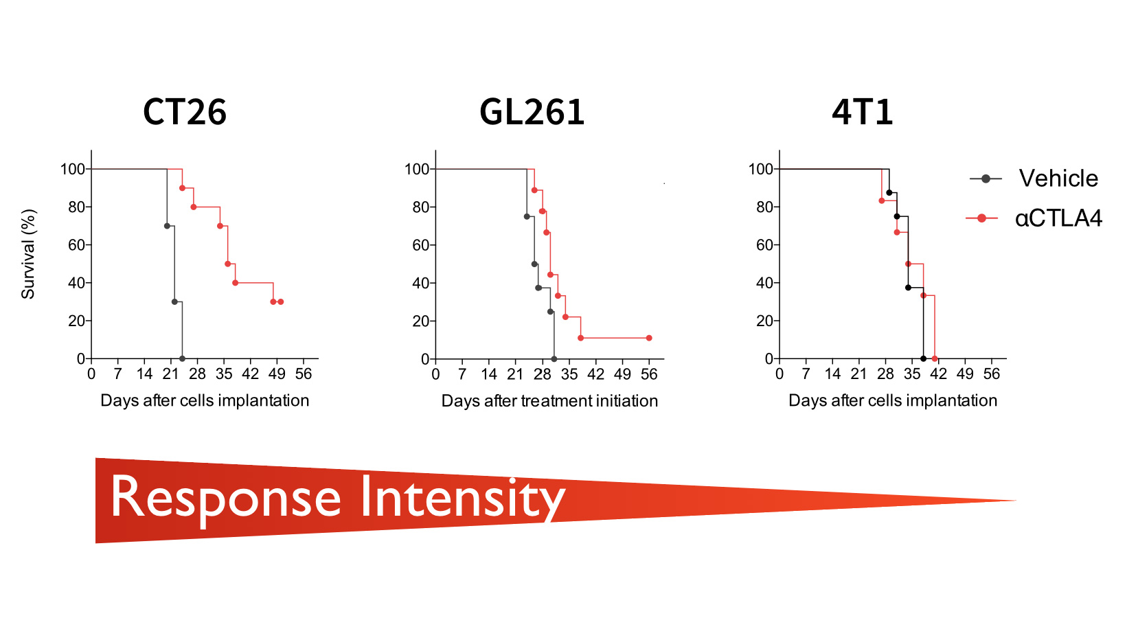
To test novel combination therapies aiming at increasing responses rates to immune checkpoint inhibitors, Explicyte has optimized a range of syngeneic mouse tumor models treated with anti-CTLA-4 or anti-PD-1/PD-L1 monoclonal antibodies. Starting with basic measurements of in vivo efficacy, our in vivo services extends to comprehensive biomarker studies, to determine the in vivo mechanism of action of novel treatments.
Our In Vivo Tumor Models Of Immune Checkpoint Blockade
- Syngeneic tumor models: tumor cells are inoculated into immunocompetent mice, so as to keep intact immunological synapse and immunoediting mechanisms.
- Responding and non-responding models to immune checkpoint inhibitors, including colorectal cancer and breast cancer models, and a bioluminescent glioblastoma model.
- Optimized treatment protocols for anti-CTLA-4 and anti PD-1/PDL-1 antibody therapies, so as to test novel combination therapies capable of leveraging anti-tumor immune response.
Assessement Of In Vivo Efficacy & Anti-Cancer Immune Response
- Standard in vivo efficacy package: Experimental groups (N=10) are monitored 3 times per week for tumor size, survival and body weight for 6 weeks.
- Additional mice for immune response profiling: Satellite mice can be included in each group and used in the course of the study to correlate in vivo effects with the activation of specific immune response, signaling pathways, etc. Explicyte offers to analyse immune biomarkers at the tumor site and in peripheral compartments (blood, spleen, lymph nodes) using quantitative multiplex technologies such as FACS or RT-qPCR.
Our Added Value
- Weekly progress reports: Our weekly reports enable clients to closely monitor the in vivo effects of their compound and eventually adjust the experimental design (treatment protocol, blood sampling, etc.).
- Longitudinal collection of serological samples : During the entire experiment, blood samples can be collected every week, allowing the monitoring of peripheral markers over time in responding and non-responding animals.
- Bioluminescent imaging: Our orthotopic tumor models (eg. intracranial glioblastoma) are based on the use of bioluminescent cell lines, thus allowing quantitative measurements of tumor progression by in vivo imaging.

