Immune Function & Modulation Assays

Macrophage Polarization & Functional Modulation Assays

Can your compound reshape macrophage phenotypes or modulate their impact on immune-cell function?

Our human macrophage assays combine M1/M2-like polarization models, phenotypic characterization and cytokine profiling with functional immune-cell co-cultures to evaluate compound-induced changes in macrophage states and associated immune responses.

Macrophage assay formats and readouts
Format
Miniaturized culture plates
Models
Human monocyte-derived M1-like or M2-like macrophages, alone or co-cultured with autologous activated PBMCs or CD4+ T cells
Macrophage phenotype
Flow cytometry markers including CD86 and CD163, where included in the panel
Cytokine profiling
IL-6, IL-10 and IL-12, among others, depending on the study design
Functional immune response
IFN-γ release as a functional readout in macrophage/immune-cell co-cultures
Reference controls
Pathway inhibitors targeting p38 MAPK, STAT3 or PI3K, and selected checkpoint antibodies, according to the study design and biological question
On request
Extended macrophage and immune-cell cytokine profiling, flow cytometry immunophenotyping, proteomics and transcriptomics
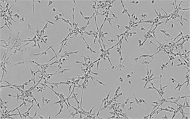
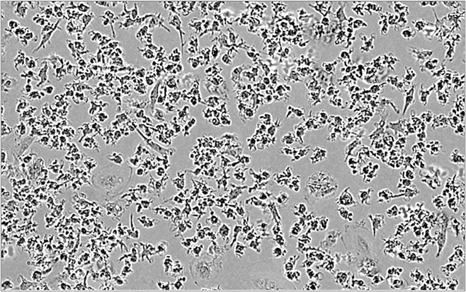
M2 macrophages M2 macrophages
M1 macrophages M1 macrophages

Macrophages adopt diverse activation states in response to environmental cues. Under defined in vitro polarization conditions, M1-like macrophages typically exhibit a more pro-inflammatory phenotype, whereas M2-like macrophages show immunoregulatory features associated with immune suppression. Our M2 suppression assay co-cultures monocyte-derived M2-like macrophages with activated CD4+ T cells or PBMCs to investigate whether test compounds modulate macrophage-associated suppression of immune-cell activity. IFN-γ release provides a functional readout of the co-culture response and is interpreted alongside macrophage cytokine profiles and, where included, phenotypic markers.

Imane Nafia, PhD, Chief Scientific Officer at Explicyte

Macrophage modulation is not simply a matter of switching between M1 and M2 states. Macrophage phenotypes are plastic and span a continuum, making it essential to distinguish effects on polarization from changes in established macrophage states, and to determine how these changes translate into immune cell function.”

Imane Nafia, PhD

Chief Scientific Officer, Explicyte

Assay principle

How our macrophage assays work
M2 Macrophage-Suppression Assay CRO immuno-oncology immunotherapy

1

Differentiate & polarize

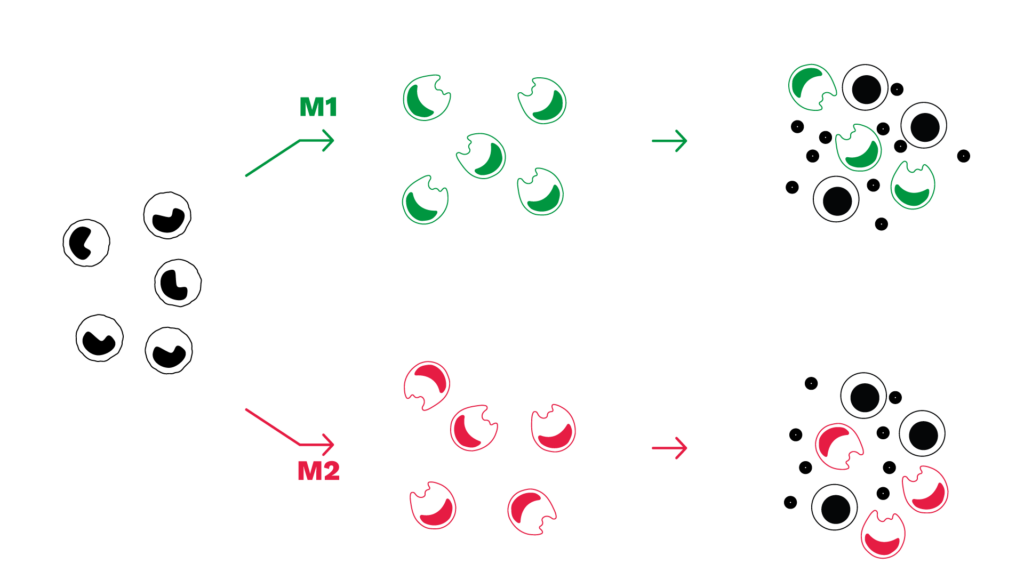
Monocytes are isolated from healthy-donor PBMCs, differentiated into macrophages, and polarized under defined in vitro conditions toward M1-like or M2-like phenotypes.

2

Co-culture with immune effector cells

M1-like or M2-like macrophages are co-cultured with autologous activated PBMCs or CD4+ T cells. Test compounds can be added during macrophage polarization, during co-culture or across both windows, depending on whether the study is designed to investigate effects on macrophage differentiation/polarization, the subsequent immune cell response, or both. Compounds can be tested individually or in combination.

3

Measure phenotype and function

IFN-γ release is quantified as a functional readout of the immune-cell response in macrophage co-cultures. In purified CD4+ T-cell co-cultures, it also serves as a surrogate of T-cell activation. Results are interpreted alongside cytokine profiles, including IL-6, IL-10 and IL-12, and macrophage phenotypic markers where assessed.

4

Interpret compound effects

Responses are evaluated against appropriate reference controls, including specific signaling pathway inhibitors and checkpoint antibodies, to contextualize compound-induced changes in macrophage phenotype and co-culture immune activity. Additional immunophenotyping or molecular profiling can be incorporated when deeper characterization is needed.

Illustrative macrophage data

Macrophage polarization and functional modulation: example results

Phenotypic and cytokine differences between M1-like and M2-like macrophages

Under defined polarization conditions, M1-like macrophages show higher CD86 and IL-6, alongside lower CD163 and IL-10, whereas M2-like macrophages display the reciprocal profile in this model.
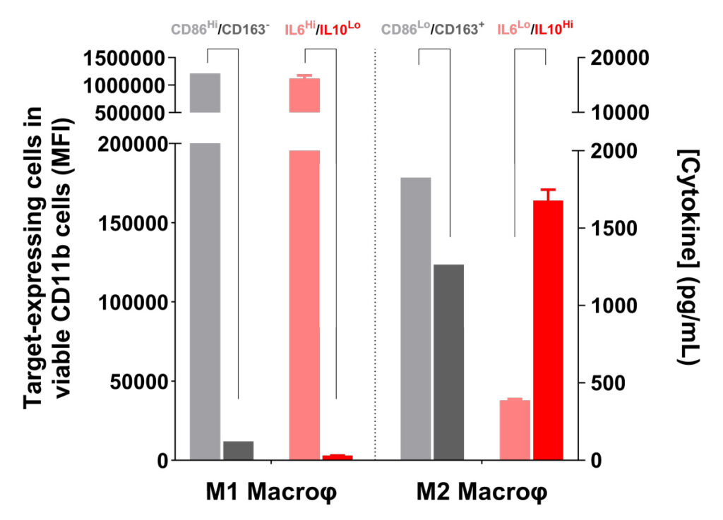
Flow cytometry marker expression and cytokine profile of M1 and M2 macrophages

Pharmacological modulation of M2-like polarization and associated immune responses

In this model, the M2-like state is characterized by an IL-6-low / IL-10-high cytokine profile. STAT3 or p38 MAPK inhibition during polarization partially shifts this profile and is associated with increased IFN-γ release in the subsequent PBMC/macrophage co-culture.

These results illustrate the assay’s ability to detect pharmacological changes in macrophage-associated cytokine profiles and their relationship with downstream immune-cell responses.
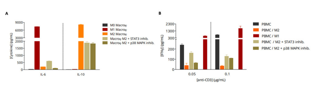
STAT3 and p38 MAPK inhibition during macrophage polarization alters cytokine profiles and co-culture IFN-γ responses.

Checkpoint blockade modulates immune responses in macrophage co-cultures

In this model, checkpoint antibodies targeting PD-1, PD-L1 or CTLA-4 increase IFN-γ release in PBMC/M2 co-cultures and further increase IFN-γ responses in PBMC/M1 co-cultures.
These results demonstrate modulation of the immune-cell response in macrophage co-cultures and support a contribution of immune checkpoint pathways to the suppressive functional context associated with M2-like macrophages in this model.
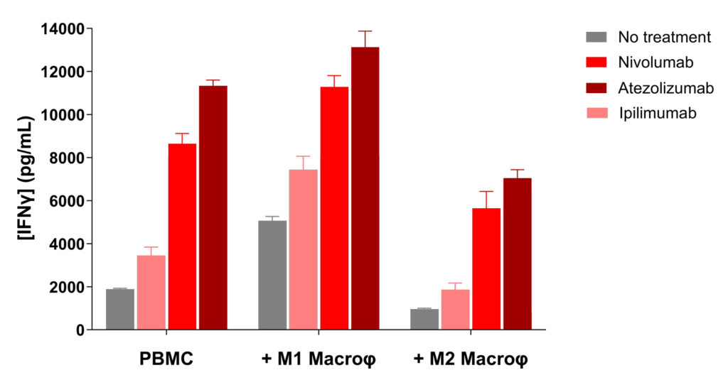
Checkpoint blockade increases IFN-γ responses in PBMC co-cultures with M1-like and M2-like macrophages

Making a difference as a preclinical CRO in oncology

Why work with Explicyte

Deep immuno-oncology experience

More than 100 in vitro campaigns over ten years, supporting 30+ peer-reviewed publications in immuno-oncology.

See our latest papers →

A dedicated PhD study director

A single PhD-level study director oversees your program from experimental planning through final report discussion.

Learn about our workflow →

Reference-anchored study design

Appropriate pathway inhibitor and checkpoint antibody controls help contextualize compound effects and support interpretation of the observed phenotype and functional response.

See our publication record →

Mechanistic readouts

IFN-γ and macrophage cytokine panels can be complemented by flow cytometry immunophenotyping, proteomics and transcriptomics to characterize cellular and molecular changes associated with compound response.

View our platform →

The Explicyte team at their Bordeaux laboratory

Paul Marteau, PharmD (study director), Imane Nafia, PhD (CSO), Loïc Cerf, MSc (COO), Alban Bessède, PhD (founder, CEO), Jean-Philippe Guégan, PhD (CTO)

Contact our team

Discuss your macrophage study

Tell us about your macrophage program — polarization, functional modulation, M2 suppression or a combination strategy — and we'll propose a fit-for-purpose study.

Explicyte (Bordeaux, France) uses your details to respond to your request. Privacy policy

Frequently asked questions

Frequently asked questions about macrophage polarization and functional assays

What do macrophage polarization and functional modulation assays measure?

These assays evaluate whether a test compound alters defined M1-like or M2-like macrophage phenotypes and how these changes are associated with immune-cell responses in co-culture. Readouts can include phenotypic markers such as CD86 and CD163, cytokine profiles including IL-6, IL-10 and IL-12, and IFN-γ as a functional co-culture readout.

Monocytes are isolated from healthy-donor PBMCs, differentiated into macrophages and polarized under defined in vitro conditions toward M1-like or M2-like phenotypes. They can then be studied alone or co-cultured with autologous activated PBMCs or CD4+ T cells.

The assay co-cultures monocyte-derived M2-like macrophages with activated CD4+ T cells or PBMCs to investigate whether test compounds modulate macrophage-associated suppression of immune-cell activity. IFN-γ provides a functional readout of the co-culture response; in purified CD4+ T-cell co-cultures, it also serves as a surrogate of T-cell activation.

Under defined polarization conditions, M1-like and M2-like macrophages show distinct phenotypic and cytokine profiles. In this model, M1-like macrophages are characterized by higher CD86 and IL-6 and lower CD163 and IL-10, whereas M2-like macrophages display the reciprocal profile. Functional co-culture readouts are interpreted alongside these macrophage-associated markers rather than using M1 and M2 as strictly binary states.

Appropriate viability controls can be incorporated to determine whether changes in macrophage phenotype or co-culture activity reflect functional modulation rather than loss of viable cells.

Reference controls may include pathway inhibitors targeting p38 MAPK, STAT3 or PI3K, as well as checkpoint antibodies such as nivolumab, atezolizumab and ipilimumab. Appropriate controls are selected according to the study design and biological question.

Yes. Test compounds can be introduced during macrophage polarization, during the subsequent immune-cell co-culture, or across both windows, depending on the biological question. Compounds can also be evaluated individually or in combination, including checkpoint antibodies and pathway modulators.

M1-like and M2-like macrophage models are reductionist in vitro systems that reproduce selected phenotypic and immunoregulatory features relevant to tumor-associated macrophage biology. They are useful for investigating TAM-relevant mechanisms and compound responses, but they do not reproduce the full heterogeneity and plasticity of macrophages within the tumor microenvironment.

Assays can be extended with broader macrophage and immune-cell cytokine profiling, flow-cytometry immunophenotyping, proteomics and transcriptomics to characterize cellular and molecular changes associated with compound response.

Macrophage polarization refers to the development of defined macrophage phenotypes under specific in vitro conditions. Compound effects can be investigated during this polarization process or, where supported by the study design, after macrophage states have been established. Distinguishing these experimental windows helps determine whether a compound affects macrophage differentiation and polarization, established macrophage phenotypes, downstream immune cell function, or a combination of these effects

Explicyte Oncology CRO logo

Capabilities

Modalities