Immune Function & Modulation Assays

Immune cell activation assay

Does your compound enhance immune-cell activation or suppress the response?

Our human immune-cell activation assays use primary PBMCs or purified T-cell populations under defined stimulation conditions to characterize compound-associated changes in cellular activation. Functional responses can be assessed through effector cytokines such as IFN-γ, IL-2 and TNF-α, together with kinetic imaging of proliferation-associated cellular responses. Reference immune modulators can be incorporated where appropriate to contextualize compound activity.

Immune cell activation assay: format & readouts
Format
96-well plate
Model
Primary human PBMCs or purified immune-cell subsets, including T cells
Activation stimulus
Anti-CD3-mediated TCR stimulation or another defined immune stimulus
Readouts

Effector cytokines including IFN-γ, IL-2 and TNF-α, with additional cytokines available according to study design

Proliferation-associated cellular responses monitored kinetically by automated live-cell imaging

Reference controls
Anti-PD-1 antibodies including nivolumab and pembrolizumab, and anti-PD-L1 atezolizumab, where appropriate to the experimental model
Additional profiling
Flow cytometry immunophenotyping, extended cytokine profiling, transcriptomics and proteomics can be incorporated to characterize cellular and molecular changes associated with compound response.
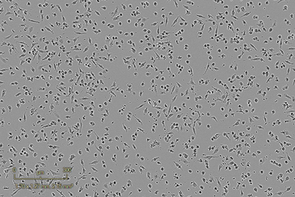
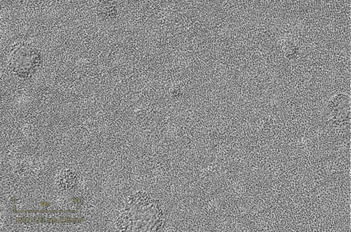
Anti-CD3 activated – 120h Anti-CD3 activated – 120h
Control – Inactivated PBMCs Control – Inactivated PBMCs

Imane Nafia, PhD, Chief Scientific Officer at Explicyte

Immune activation is not simply an on/off switch. The strength, timing and quality of the response all matter. A useful functional assay therefore needs to show not only whether a candidate increases or suppresses activation, but how it reshapes the response across complementary cellular and cytokine readouts. This helps place compound activity in a biological context that is relevant to immunotherapy development.

Imane Nafia, PhD

Chief Scientific Officer, Explicyte

Assay principle

From immune-cell stimulation to functional readouts
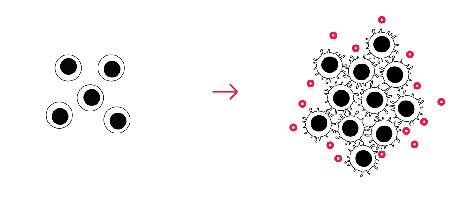
Immune cell activation assay workflow — PBMC/T cell stimulation, live-cell imaging, and cytokine quantification

1

Stimulate immune cells

PBMCs or purified T-cell populations are exposed to anti-CD3-mediated TCR stimulation or another defined immune stimulus according to the biological question.

2

Track the cellular response kinetically

Automated live-cell imaging is used to monitor proliferation-associated changes and cellular clustering over time, providing a kinetic view of the response to stimulation and compound treatment.

3

Quantify effector cytokines

IFN-γ, IL-2, TNF-α and additional cytokines can be quantified to characterize the functional immune response and its modulation by the test compound.

4

Add complementary biological characterization

Flow cytometry immunophenotyping, broader cytokine profiling, proteomics or transcriptomics can be integrated to characterize cellular and molecular changes associated with the observed functional response.

illustrative immune cell activation data

Immune activation and suppression: representative results

Anti-CD3 induces dose- and time-dependent effector cytokine responses

Anti-CD3 stimulation induces TNF-α release within 24 hours, while IFN-γ and IL-2 increase with stimulation strength and exposure time. Together, these cytokine profiles provide complementary functional readouts of the evolving immune response.
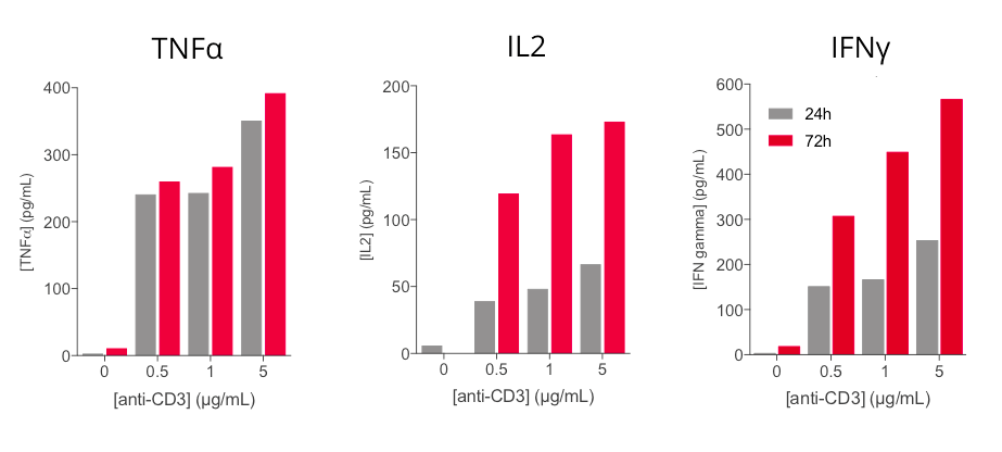
Line graphs of TNF-α, IL-2, and IFN-γ release from human PBMCs across anti-CD3 dose and time

Anti-PD-1 antibodies enhance anti-CD3-induced IFN-γ responses

Increasing concentrations of nivolumab or pembrolizumab enhance IFN-γ release in anti-CD3-activated PBMCs in a dose-dependent manner. These data illustrate the assay’s ability to detect checkpoint inhibitor-associated enhancement of the immune response.
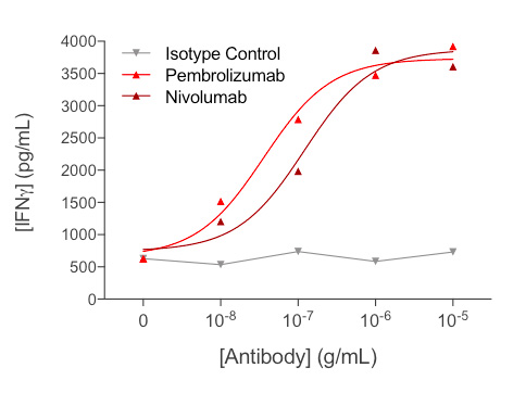
Dose-response of IFN-γ release from anti-CD3-activated PBMCs treated with nivolumab or pembrolizumab

L-kynurenine suppresses activated T-cell responses

In primary CD4+ T cells stimulated with anti-CD3, L-kynurenine reduces TNF-α and IFN-γ levels in a concentration-dependent manner after 72 hours.

These data illustrate the assay’s ability to characterize both enhancement and suppression of activated T-cell responses.
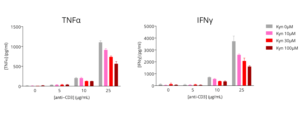
Dose-dependent reduction of TNF-α and IFN-γ in anti-CD3-stimulated CD4+ T cells with L-kynurenine

Making a difference as a preclinical CRO in oncology

Human-primary data, read in both directions and in depth

A dedicated study director

A PhD-level study director oversees the program from experimental planning through final data interpretation and report discussion.

More about us →

Human primary cells

Studies use primary human PBMCs or purified immune-cell populations to capture biologically relevant responses and, where needed, donor-to-donor variability.

About our cell-based assays →

Functional modulation in both directions

Appropriate reference immune modulators can be incorporated to contextualize compound-associated enhancement or suppression of the activated immune response.

Discuss your controls →

Kinetic and complementary readouts

Cellular responses can be followed over time and complemented with cytokine, phenotypic and molecular profiling to provide biological context beyond a single endpoint.

See our platforms →

The Explicyte team at their Bordeaux laboratory

Paul Marteau, PharmD (study director), Imane Nafia, PhD (CSO), Loïc Cerf, MSc (COO), Alban Bessede, PhD (founder, CEO), Jean-Philippe Guégan, PhD (CTO)

Contact our team

Discuss your immune cell activation project

Tell us your compound or modality, the biological question, preferred cell model and desired readouts. We’ll propose the donor strategy, stimulation conditions, controls, dose range and analysis plan.

Explicyte (Bordeaux, France) uses your details to respond to your request. Privacy policy

Frequently asked questions

Frequently asked questions about immune cell activation assays

What does the immune cell activation assay measure?

It evaluates whether a compound modulates the activation of primary human immune cells following defined stimulation. Effector cytokines provide functional readouts, while live-cell imaging can capture proliferation-associated cellular responses and clustering over time, allowing both enhancement and suppression to be characterized.

Primary human PBMCs, or a purified subset such as T cells, depending on the question. Working in primary cells means the response reflects donor biology rather than an immortalized line.

Yes. PBMC or purified T-cell studies can be designed across multiple independent donors to assess reproducibility and inter-donor variability. Donor number, treatment conditions and replication are defined according to the study objective.

Immune-cell activation is characterized using complementary functional readouts. Effector cytokines such as IFN-γ, IL-2 and TNF-α are quantified at defined timepoints, while automated live-cell imaging can provide kinetic information on proliferation-associated cellular responses and clustering.

IFN-γ, IL-2 and TNF-α are standard readouts. IL-2 and IFN-γ are key measures of T-cell activation in this assay, while TNF-α can provide an earlier inflammatory readout. The optimal sampling time is defined according to the stimulus and study design.

Reference controls can include anti-PD-1 antibodies such as nivolumab or pembrolizumab, and the anti-PD-L1 antibody atezolizumab. Controls are selected according to the study design and run under the same assay conditions as the test compound, providing a relevant benchmark for interpreting potentiating or suppressive effects on immune-cell activation.

Yes. The assay can detect responses in both directions. In representative data, checkpoint inhibition increases IFN-γ output in anti-CD3-activated PBMCs, whereas L-kynurenine produces a concentration-dependent reduction in cytokine responses in activated primary CD4+ T cells.

Additional analyses can include flow cytometry immunophenotyping, broader cytokine profiling, proteomics and transcriptomics. These readouts can help characterize cellular and molecular changes associated with compound response and support mechanistic investigation.

Yes. Compounds and antibodies can be run alone and in combination with anti-CD3, which is how checkpoint inhibitors are profiled — reading the added effect on cytokine output and proliferation-associated cellular responses against the anti-CD3 baseline.

Explicyte Oncology CRO logo

Capabilities

Modalities