Immune Function & Modulation Assays

Germinal Center Reaction Assay

Does your compound modulate Tfh–B-cell interactions relevant to germinal-center biology and tertiary lymphoid structures (TLS)?

Our human tonsil-derived assay combines autologous T follicular helper (Tfh) cells and memory B cells to characterize compound effects on B-cell function and Tfh-supported responses. IgG secretion provides a primary functional readout, while complementary immunophenotyping and molecular profiling can characterize changes in B-cell differentiation, phenotype and associated immune programs.

Germinal center reaction assay: format & readouts
Format
96-well plate co-culture
Model
Autologous human tonsil-derived T follicular helper cells (CD4+/CXCR5+) and memory B cells (Bmem, CD19+/CD27+)
Activation
Staphylococcal enterotoxin B (SEB)
Functional readout
Secreted IgG as a measure of antibody secretion
Response
Detects increases or decreases in IgG output following compound treatment
Cellular phenotyping
Flow cytometry can be incorporated to characterize Tfh and B-cell phenotypes and compound-associated changes in B-cell differentiation or activation, according to the study design
Reference controls
SEB; checkpoint inhibitors including nivolumab, atezolizumab and ipilimumab; and the CD40-blocking antibody bleselumab, selected according to the biological question
Additional profiling
Extended cytokine profiling and complementary proteomic, transcriptomic or signaling analyses to characterize cellular and molecular changes associated with compound response
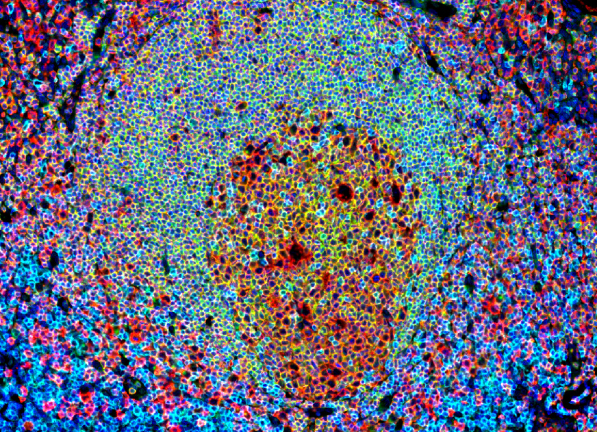
Immunohistofluorescence field of a human tonsil section illustrating the germinal center in a lymphoid structure.

Human tonsil tissue provides the cellular foundation of the assay
Immunohistofluorescence of a human tonsil illustrates a germinal center within a lymphoid follicle and the spatial organization of Tfh and B-cell populations. Human tonsils provide a rich source of the autologous Tfh and memory B cells used in our functional co-culture assay.

Imane Nafia, PhD, Chief Scientific Officer at Explicyte

Germinal-center biology is not only about which immune cells are present, but how they function together. By probing Tfh–B-cell interactions, our assay provides a controlled way to investigate whether a candidate modulates a key component of the humoral immune response. IgG secretion provides a functional endpoint, while complementary cellular and molecular profiling can characterize the biological changes associated with that response and add a functional dimension to TLS-related research.

Imane Nafia, PhD

Chief Scientific Officer, Explicyte

Assay principle

From Tfh–B-cell co-culture to functional and phenotypic readouts
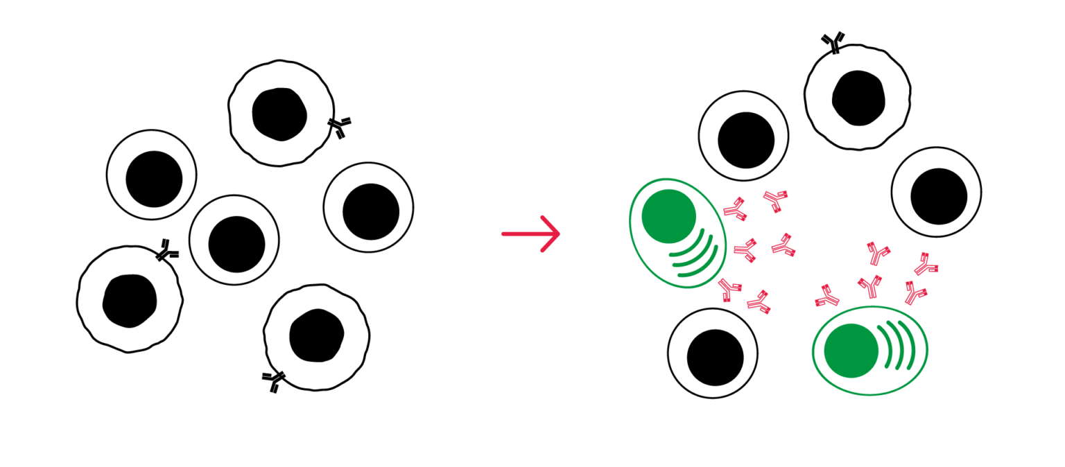
Germinal center reaction assay workflow using human Tfh and memory B-cell co-culture with functional and phenotypic readouts

1

Isolate and co-culture

Autologous Tfh cells and memory B cells are isolated from human tonsil and co-cultured with the test compound, alone or in combination with selected immune modulators according to study design.

2

Activate the co-culture

SEB provides an activation stimulus that establishes a functional Tfh–B-cell response against which compound-associated changes can be evaluated.

3

Measure the functional response

Secreted IgG is quantified in the supernatant as a functional measure of antibody secretion, enabling detection of compound-associated increases or decreases in the response.

4

Characterize cellular and molecular changes

Flow cytometry immunophenotyping and complementary cytokine, proteomic or transcriptomic analyses can be incorporated to characterize changes in Tfh and B-cell phenotype, differentiation and associated biological programs.

illustrative Germinal center reaction data

Tfh–B-cell co-culture: example results

Isolation and phenotypic characterization of Tfh and memory B cells

Freshly isolated Tfh and memory B cells were characterized by multiparameter flow cytometry before and after isolation. Tfh cells were identified as CD4+/CXCR5+, while memory B cells were identified as CD19+/CD27+, confirming enrichment of the intended cell populations used in the functional co-culture assay.
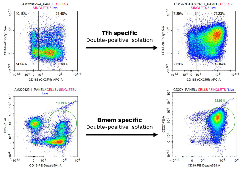
Flow cytometry characterization of Tfh (CD4+/CXCR5+) and memory B (CD19+/CD27+) cells before and after isolation

Reference immune modulators increase or decrease IgG secretion

Tfh and memory B cells were co-cultured under SEB activation, and secreted IgG was quantified in the supernatant on day 7. The checkpoint inhibitors nivolumab, atezolizumab and ipilimumab increased IgG secretion, whereas the CD40-blocking antibody bleselumab decreased it.
These results illustrate the assay’s ability to detect both increases and decreases in Tfh-supported B-cell responses using reference immune modulators with distinct mechanisms of action.
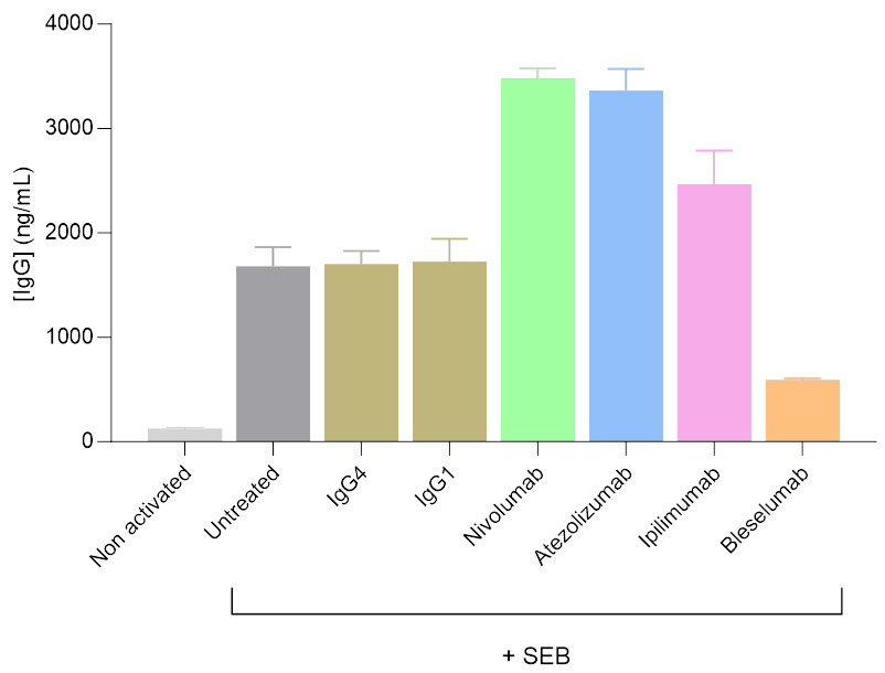
IgG secretion from an SEB-activated Tfh/Bmem co-culture modulated by anti-PD-1, anti-PD-L1, anti-CTLA-4 and anti-CD40 antibodies

Concentration-dependent suppression of IgG secretion by kynurenine

In the same SEB-activated Tfh–B-cell co-culture, kynurenine induced a concentration-dependent decrease in IgG secretion, both in the presence and absence of anti-PD-1 treatment with nivolumab.
These results illustrate the assay’s ability to quantify concentration-dependent suppression of the functional response and to evaluate how compound-associated effects behave in a checkpoint-modulated context.
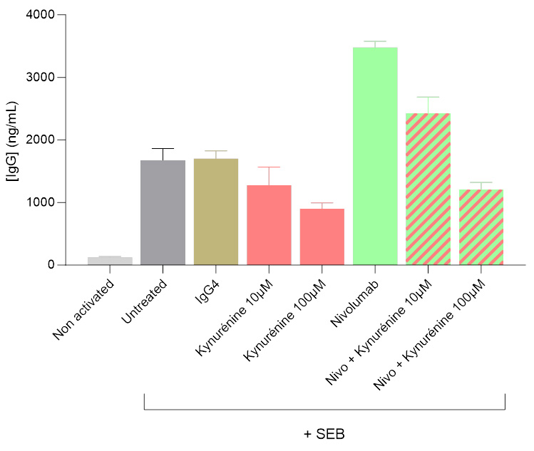
Dose-dependent decrease in IgG secretion from a Tfh/Bmem co-culture treated with kynurenine, with and without anti-PD-1

Tfh–B-cell interactions shape the cytokine and B-cell-associated soluble-factor response

Memory B cells isolated from human tonsil were cultured either under IL-21/IL-4/CD40L stimulation, reproducing key signals delivered by Tfh cells (top panel), or in co-culture with autologous Tfh cells under SEB activation (bottom panel).

Supernatants were analyzed using a multiplex approach for IFN-γ, TNF-α, TNF-β, IL-4, IL-6, IL-10, BAFF and APRIL. The two conditions generate distinct soluble-factor profiles, illustrating that direct Tfh–B-cell interactions provide an additional level of regulation beyond defined Tfh-mimetic stimulation.

These data demonstrate the assay’s ability to characterize how Tfh–B-cell cross-talk modulates cytokine and B-cell-associated responses, complementing IgG secretion with a broader functional view of germinal-center-related biology.
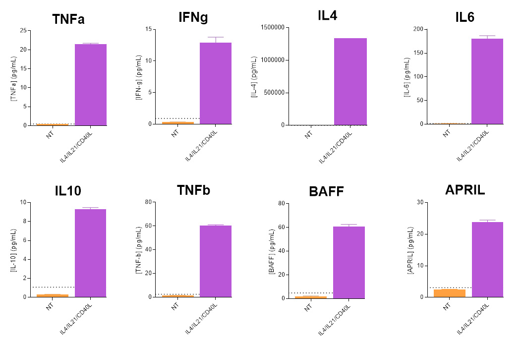
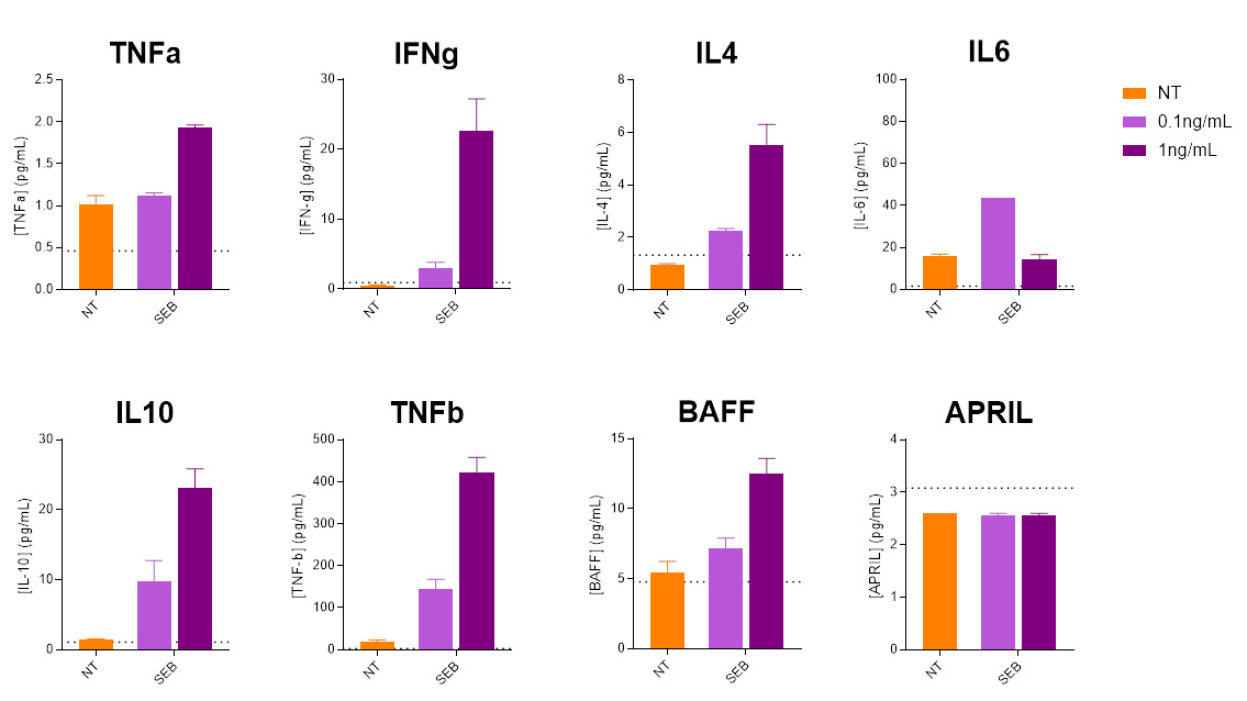
Cytokine profiling of activated human tonsil-derived memory B cells Cytokine profiling of human Tfh and memory B-cell co-cultures

Making a difference as a preclinical CRO in oncology

Why work with Explicyte for germinal-center and B-cell functional assays

100+ in vitro campaigns

More than 100 in vitro campaigns over ten years, with a dedicated PhD-level study director overseeing each program from experimental planning through final report discussion.

More about us →

Depth in TLS and B cell biology

Co-authors of peer-reviewed publications on the role of TLS in immunotherapy response, we have also developed ISO-certified pathology services for TLS scoring in IO trials.

Explore TLS detection & scoring →

Reference-anchored functional assessment

Reference immune modulators can be incorporated to contextualize compound-associated increases or decreases in functional and cellular readouts and support interpretation of response magnitude and direction.

Discuss your controls →

Extend beyond the primary functional readout

Flow cytometry immunophenotyping, cytokine profiling, proteomics and transcriptomics can be incorporated to characterize cellular and molecular changes associated with compound response.

See our platforms →

The Explicyte team at their Bordeaux laboratory

Paul Marteau, PharmD (study director), Imane Nafia, PhD (CSO), Loïc Cerf, MSc (COO), Alban Bessede, PhD (founder, CEO), Jean-Philippe Guégan, PhD (CTO)

Contact our team

Discuss your germinal center program

Tell us the question you need the germinal center reaction assay to answer — which compounds, which controls, and the readouts beyond IgG you want on the same co-culture. A PhD-level study director will scope it with you.

Explicyte (Bordeaux, France) uses your details to respond to your request. Privacy policy

Frequently asked questions

Frequently asked questions about germinal center reaction and Tfh–B-cell assays

What does the germinal center reaction assay measure?

The assay models a key functional interaction between human tonsil-derived T follicular helper (Tfh) cells and memory B cells relevant to germinal-center biology. It can be used to evaluate how a test compound modulates Tfh-supported B-cell responses through functional readouts such as antibody secretion, together with cellular phenotyping and complementary cytokine or molecular profiling.

The assay uses autologous T follicular helper cells and memory B cells isolated from human tonsil. Tfh cells are characterized as CD4+/CXCR5+, while memory B cells are characterized as CD19+/CD27+. Human tonsil provides a rich source of these interacting immune-cell populations for functional co-culture studies.

Secreted IgG provides a primary functional measure of antibody secretion. Flow cytometry immunophenotyping can be incorporated to characterize Tfh and B-cell phenotypes and compound-associated changes in B-cell activation or differentiation. Extended cytokine profiling and complementary proteomic, transcriptomic or signaling analyses can also be integrated according to the study design.

Yes. Flow cytometry can be incorporated to characterize changes in B-cell phenotype, activation and differentiation alongside the functional response. The markers and cell populations assessed are selected according to the biological question and the expected effect of the test compound.

Multiplex cytokine profiling can provide an additional functional layer beyond IgG secretion. Depending on the study design, analytes can include IFN-γ, TNF-α, TNF-β, IL-4, IL-6, IL-10, BAFF and APRIL. Profiling B-cell cultures and Tfh–B-cell co-cultures under defined experimental conditions can help characterize cytokine and soluble-factor responses associated with B-cell activation and Tfh–B-cell interactions.

Secreted IgG is quantified in the co-culture supernatant and used as a functional readout of the Tfh-supported B-cell response. Changes in IgG secretion can reveal compound-associated enhancement or suppression of antibody production and are interpreted alongside other cellular or molecular readouts when included.

SEB is used to establish an activated Tfh–B-cell co-culture response. Reference immune modulators can include checkpoint inhibitors such as nivolumab, atezolizumab and ipilimumab, as well as the CD40-blocking antibody bleselumab. Appropriate controls are selected according to the study design and biological question.

Yes. In representative data, checkpoint inhibitors increase IgG secretion, whereas CD40 blockade with bleselumab decreases it. Kynurenine also produces a concentration-dependent reduction in IgG secretion. These responses illustrate the assay’s ability to detect both enhancement and suppression and to quantify graded compound effects.

Appropriate viability controls can be incorporated to determine whether reduced IgG secretion or changes in other functional readouts reflect modulation of the Tfh–B-cell response rather than loss of viable cells. Cellular phenotyping can provide additional context when needed.

Tfh–B-cell interactions are a key component of germinal-center biology and are also relevant to organized immune responses within mature tertiary lymphoid structures. This in vitro assay provides a controlled system for investigating one functional component of TLS-associated biology, but it does not reproduce the full cellular and spatial complexity of a tertiary lymphoid structure.

The germinal center reaction assay is an in vitro functional Tfh–B-cell co-culture used to investigate compound-associated changes in defined cellular responses. TLS detection and scoring is a separate tissue-based approach that identifies and characterizes tertiary lymphoid structures in patient samples, including their organization and maturity. The two approaches are complementary: one provides functional information, while the other characterizes TLS biology directly in tissue.

Yes. Test compounds can be evaluated alone or in combination with selected immune modulators or other relevant agents according to the study design. This allows compound-associated effects to be assessed in different immunological contexts, including checkpoint-modulated conditions.

Studies can be extended with flow cytometry immunophenotyping, broader cytokine profiling, proteomics, transcriptomics and selected signaling analyses. These complementary readouts can help characterize cellular and molecular changes associated with compound response beyond the primary IgG functional endpoint.

Explicyte Oncology CRO logo

Capabilities

Modalities