Immune Function & Modulation Assays

Cell Migration & Chemotaxis Assays

Does your compound modulate immune-cell recruitment or tumor-cell migration under defined chemotactic and microenvironmental conditions?

Our kinetic transwell migration assays combine defined chemotactic conditions with live-cell imaging to quantify immune-cell chemotaxis and tumor-cell migration over time. Primary immune cells and selected cancer-cell models can be used to evaluate compound-associated enhancement or inhibition of migration, with complementary phenotypic and molecular profiling available to characterize the biological response.

Cell migration assay: format & readouts

Format
96-well transwell plates with pore-based inserts, with extracellular matrix coating where appropriate.
Models
– Primary or established immune-cell models, including T cells and neutrophils, with additional immune-cell subsets available according to study design – Selected cancer-cell models for tumor-cell migration studies
Migration readout
Cell migration from the upper to the lower chamber quantified kinetically by live-cell imaging over 6–48 hours.
Chemotactic conditions
CXCL12 (SDF-1α) for T cells; CXCL8 (IL-8), CXCL1 and fMLP for neutrophils; serum-based gradients for tumor-cell migration. Additional chemoattractants or stimuli can be incorporated according to the biological question.
Compound evaluation
Test compounds can be assessed for their ability to promote or inhibit migration, or to modulate the cellular response to a defined chemotactic stimulus.
Additional profiling
Flow cytometry immunophenotyping, cytokine profiling, transcriptomics and proteomics can be incorporated to characterize cellular and molecular changes associated with the migration phenotype.

Real-time chemotactic migration of immortalized T cells toward SDF1α in a 96-well plate. Transmembrane migration quantified on the IncuCyte® live-cell analysis system.

Imane Nafia, PhD, Chief Scientific Officer at Explicyte

Migration is more than movement. It is a biological response shaped by the cellular model, chemotactic signals and microenvironmental context. From immune-cell recruitment to tumor-cell migration, we investigate how cells respond under defined conditions, how a compound modulates that response, and how these effects can inform proof-of-concept and mechanistic studies.

Imane Nafia, PhD

Chief Scientific Officer, Explicyte

Assay principle

How our cell migration and chemotaxis assays work
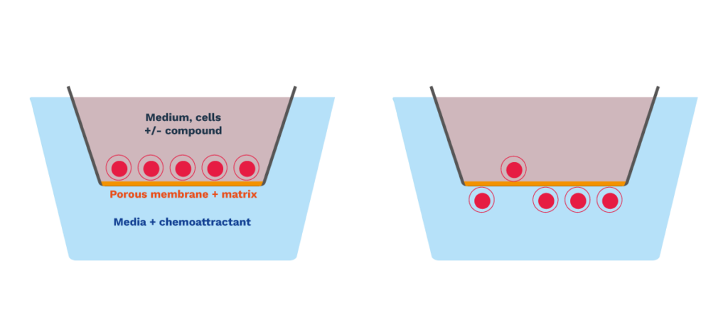
immune cell migration assays chemotaxis cancer immunotherapy t cells and neutrophils

1

Prepare and seed the cells

Primary immune cells or selected cancer-cell models are prepared and seeded into the upper chamber of a pore-based transwell insert. Extracellular matrix coating can be incorporated where appropriate to the biological model.

2

Establish the migration condition

A defined chemotactic or migration-promoting condition is established in the lower chamber. Test compounds can be evaluated for their ability to promote or inhibit migration, or to modulate the response to the selected stimulus.

3

Track migration kinetically

Migration toward the lower chamber is quantified by live-cell imaging over a 6–48-hour window, providing a time-resolved profile of migration onset, rate and magnitude rather than a single terminal endpoint.

4

Add complementary biological readouts

Flow cytometry immunophenotyping, cytokine profiling, transcriptomics or proteomics can be integrated to characterize cellular and molecular changes associated with the migration phenotype.

Illustrative CELL MIGRATION data

T-cell, neutrophil and tumor-cell migration: representative results

Dose-dependent T-cell chemotaxis is inhibited by CXCR4 blockade

Primary human T-cell migration is induced in a concentration-dependent manner by CXCL12 (SDF-1α). CXCL12-driven migration is inhibited by the CXCR4 antagonist AMD3100, illustrating the assay’s ability to detect pathway-dependent modulation of T-cell chemotaxis.
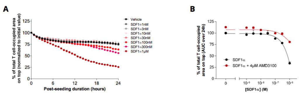
T-cell chemotaxis assay: SDF1α dose-response reversed by the CXCR4 inhibitor AMD-3100

Neutrophil chemotaxis toward CXCL8 and CXCL1 is modulated by CXCR2 antagonism

Freshly isolated human neutrophils show concentration-dependent migration toward CXCL8 (IL-8) and CXCL1. Treatment with a CXCR2 antagonist partially inhibits migration induced by both chemokines, illustrating pharmacological modulation of neutrophil chemotaxis.
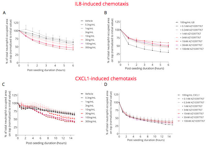
Neutrophil chemotaxis assay: IL-8 and CXCL1 dose-response blocked by CXCR2 antagonism

Serum-induced sarcoma cell migration is enhanced by fibronectin

Sarcoma cell migration was monitored by live-cell imaging over 48 hours in a transwell system. NucLight fluorescence was quantified in the upper and lower chambers and used to calculate migration rate.Higher serum conditions promoted sarcoma cell migration, while fibronectin further increased the migratory response.

These data illustrate the assay’s ability to quantify kinetic tumor-cell migration and evaluate how extracellular-matrix components modulate the response to migration-promoting conditions.
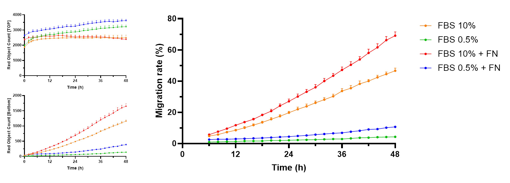
Serum-induced sarcoma cell migration and modulatory effects of fibronectin.

Making a difference as a preclinical CRO in oncology

Why work with Explicyte

The right model for the biological question

We select the cell population and assay configuration that best reflect the biology being tested, from primary T cells and neutrophils to appropriate tumor-cell models.

Our expertise in cell-based assays →

Ten years of functional oncology and immuno-oncology assays

More than 100 in vitro campaigns over the past decade, supported by complementary technologies to characterize cellular responses beyond migration alone.

View our platform →

Published, peer-reviewed science

30+ papers in key immuno-oncology journals. Assay performance is assessed using appropriate reference chemoattractants, pathway inhibitors and experimental controls.

See our latest papers →

One study director, end to end

A dedicated PhD-level study director takes your program from experimental plan to final-report discussion, with a fit-for-purpose proposal built around your objective.

Discuss your project →

The Explicyte team at their Bordeaux laboratory

Paul Marteau, PharmD (study director), Imane Nafia, PhD (CSO), Loïc Cerf, MSc (COO), Alban Bessède, PhD (founder, CEO), Jean-Philippe Guégan, PhD (CTO)

Contact our team

Discuss your migration study

Tell us about your compound and the migration question you need answered — recruiting effector cells, or blocking cancer-cell or suppressive-cell migration. We'll come back with a fit-for-purpose proposal and a PhD-level study director.

Explicyte (Bordeaux, France) uses your details to respond to your request. Privacy policy

Frequently asked questions

Frequently asked questions about cell migration and chemotaxis assays

What does a cell migration or chemotaxis assay measure?

Cell migration assays quantify the movement of cells under defined experimental conditions. In chemotaxis assays, migration occurs in response to a defined chemical gradient. Explicyte uses kinetic transwell assays to evaluate whether test compounds promote or inhibit immune-cell chemotaxis, tumor-cell migration, or the cellular response to a selected migration-promoting stimulus.

Cell migration is the broader process of cellular movement, whereas chemotaxis specifically refers to directed migration in response to a chemical gradient. Depending on the biological question, our assays can model immune-cell chemotaxis toward defined chemokines or tumor-cell migration under serum- or extracellular-matrix-modulated conditions.

Primary immune-cell populations can include T cells and freshly isolated neutrophils, with additional immune-cell subsets available according to study design. Selected established cancer-cell models can also be used to investigate tumor-cell migration. The cell model is selected according to the biological question and the expected mechanism of the test compound.

Cells are placed in the upper chamber of a pore-based transwell system, while a migration-promoting condition is established in the lower chamber. Live-cell imaging is then used to follow cell movement over time, typically across a 6–48-hour window depending on the model. This provides a kinetic profile of migration rather than a single terminal measurement.

Representative conditions include CXCL12 (SDF-1α) for T-cell chemotaxis and CXCL8 (IL-8), CXCL1 or fMLP for neutrophil migration. Serum-based gradients can be used for selected tumor-cell migration models. Other chemoattractants, stimuli or migration-promoting conditions can be incorporated according to the biological question.

Yes. Test compounds can be evaluated for their ability to increase migration, inhibit migration toward a defined chemoattractant, or modulate the response to an established migration-promoting condition. Appropriate reference controls and inhibitors can be incorporated according to the pathway or mechanism being investigated.

Yes. In addition to immune-cell chemotaxis, selected tumor-cell models can be evaluated in transwell migration assays. For example, serum-driven sarcoma-cell migration can be quantified kinetically and used to investigate how test compounds or extracellular-matrix conditions modify the migratory response.

Transwell inserts can be coated with extracellular-matrix components when relevant to the biological model. This allows the assay to evaluate how matrix-associated conditions influence cell migration. In representative sarcoma data, fibronectin enhanced migration under serum-driven conditions, illustrating how extracellular-matrix components can modulate the migratory phenotype.

Appropriate viability controls can be incorporated to determine whether reduced migration reflects a change in migratory behavior rather than loss of viable cells. Additional phenotypic or functional readouts can also be included when required to contextualize the observed response.

Migration assays can be complemented with flow cytometry immunophenotyping, cytokine profiling, transcriptomics and proteomics. These additional analyses can help characterize cellular and molecular changes associated with the migration phenotype and provide biological context beyond the migration readout alone.

Live-cell imaging provides a time-resolved migration profile rather than a single endpoint measurement. This makes it possible to compare migration onset, rate, magnitude and plateau across experimental conditions and can reveal differences in migration kinetics that may be missed by a terminal readout.

Explicyte Oncology CRO logo

Capabilities

Modalities