TUMOR ORGANOIDS AND FRAGMENTS

Patient-Derived 3D Tumor Models: Immune-Containing Organoids and Tumor Fragments

Explicyte establishes short-term 3D tumor models directly from fresh patient tumor tissue, retaining endogenous immune populations over the experimental window for functional immunotherapy testing.

By capturing patient-specific tumor features and immune context, these models provide a human-relevant in vitro platform to investigate treatment response and immune modulation in patient-derived tissue, and generate translational evidence that complements and strengthens conventional preclinical models.

TUMOR ORGANOIDS AND FRAGMENTS

Why patient-derived, immune-containing 3D models?

Patient-derived 3D tumor models are established directly from fresh patient tumor tissue as early-passage organoids or intact tumor fragments. Depending on the model and experimental window, they retain endogenous immune populations alongside patient-specific tumor features.

Custom 3D models

Through our biobank network, we source fresh tumor tissue from surgical resections or biopsies and establish short-term patient-derived 3D models tailored to the program — from early-passage organoids to intact tumor fragments.

Translational relevance

Both formats preserve patient-specific tumor characteristics and heterogeneity. Early organoids can retain a fraction of the endogenous immune compartment during the experimental window, while intact tumor fragments additionally preserve tissue architecture and spatial relationships within the TME.

From response to mechanism

Beyond supernatant analysis and kinetic functional readouts, we integrate spatial biology — digital pathology and single-cell spatial transcriptomics — to investigate which cells respond, where responses occur within the tissue, and which cellular programs are associated with treatment response.
Imane Nafia, PhD, Chief Scientific Officer at Explicyte

For immunotherapy, the biology matters. Patient-derived 3D models retain key elements of the patient’s tumor and immune context, allowing treatments to be tested in a setting that is closer to the biology they are intended to modulate.

Early organoids provide a short-term window into functional immune responses, while tumor fragments preserve the native tissue architecture and spatial organization of the TME.

Combined with functional, cytokine and spatial readouts, these models allow us to see not only whether a treatment works, but which immune cells respond, where the response occurs, and which cellular programs are engaged.

Imane Nafia, PhD

Chief Scientific Officer, Explicyte

Early tumor organoids

Immune-containing patient-derived tumor organoids (PDTOs)

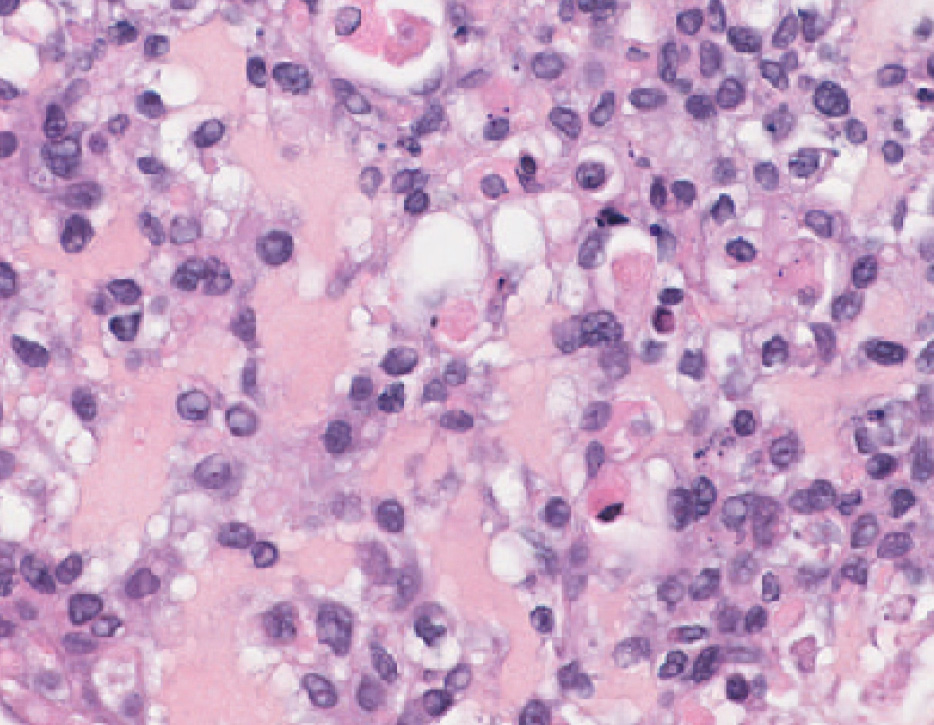
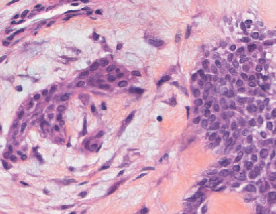
Short-term patient-derived tumor organoids established directly from fresh tumor tissue with no or minimal passage, retaining a fraction of endogenous immune populations during the experimental window.

At a glance

Core features of early PDTOs
Origin
Fresh patient tumor tissue, no/minimal passage
Immune content
Fraction of endogenous immune populations retained
Experimental window
~7 days
Readouts
Flow cytometry · cytokines · functional response kinetics
Best for
Short-term & early immunotherapy testing in patient-derived 3D cultures
Organoid — 1 week Organoid — 1 week
TNBC patient tumor TNBC patient tumor

Preserved immune fraction and IL2-sustained CD8 persistence

Early immune-containing TNBC organoids retain a measurable fraction of T cells relative to the parent tumor. Under IL2 supplementation, the CD8 compartment persists through day 7, where the untreated condition declines, demonstrating dynamic, culture-dependent changes in the retained immune compartment rather than a static endpoint measurement.

Functional immune modulation in early immune-containing TNBC PDTOs

Early immune-containing TNBC PDTOs respond to immune stimulation and checkpoint modulation. Anti-CD3 stimulation and nivolumab modulate the CD45, CD3, CD4 and CD8 compartments, providing functional readouts of immune cell engagement in patient-derived 3D cultures.

PDTF MODELS

Patient-Derived Tumor Fragments

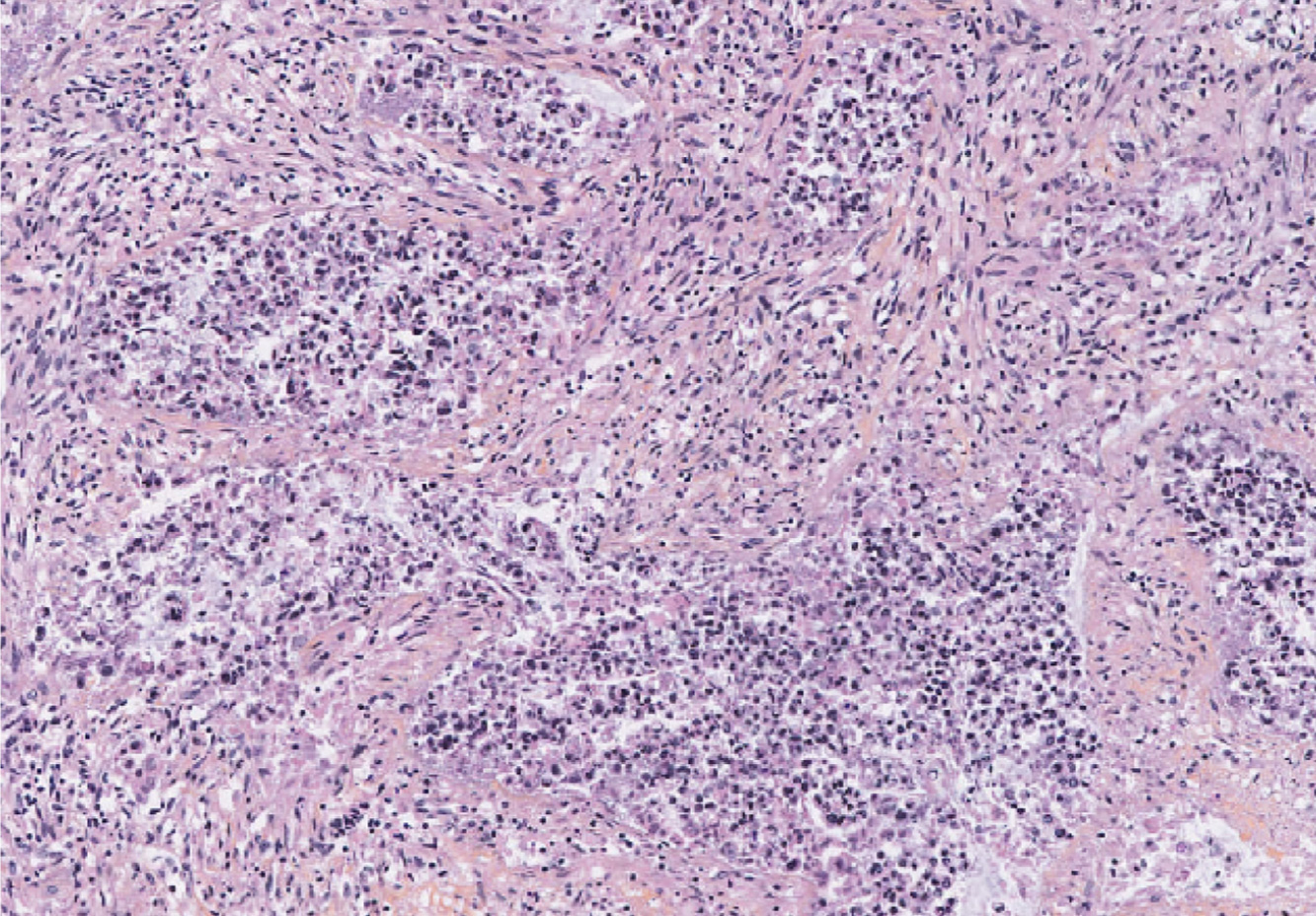
A highly patient-proximal ex vivo model that retains intact tumor tissue architecture and the endogenous immune infiltrate in situ, enabling functional immunotherapy testing directly on patient-derived tissue.

At a glance

Core features of PDTFs
Origin
Fresh patient tumor tissue
Immune content
Endogenous immune infiltrate retained in situ
Architecture
Native tumor and TME architecture preserved
Experimental window
2–5 days
Readouts
Flow cytometry · cytokines · digital pathology · spatial profiling
Best for
Immunotherapy response and biomarker evaluation in intact patient-derived tumor tissue before clinical trial initiation
Colorectal cancer (CRC) patient-derived tumor fragment (PDTF) after 2 days of anti-CD3 treatment

2-day colorectal cancer fragment, treated with anti-CD3

Quantifying the native immune infiltrate

Flow cytometry of a CRC fragment resolves the native immune infiltrate directly from patient tissue, including T cells (CD8, CD4), myeloid cells (CD11b, CD11c), NK (CD16), and B cells (CD19), with each population quantified as a fraction of viable CD45⁺ cells before treatment.
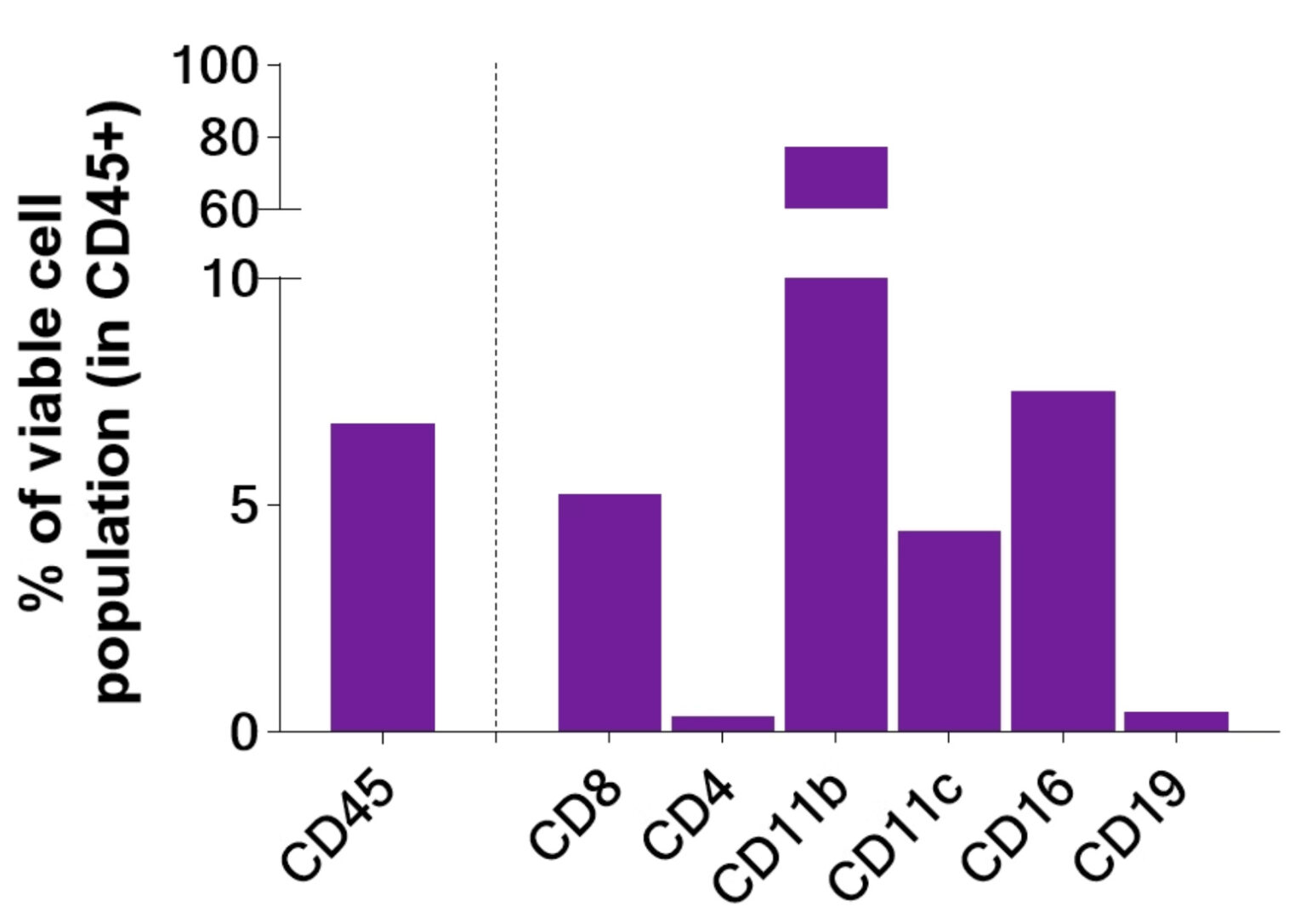
Flow cytometry of a CRC patient-derived tumor fragment resolving the native immune infiltrate: T cells (CD8, CD4), myeloid cells (CD11b, CD11c), NK cells (CD16) and B cells (CD19), quantified as fractions of viable CD45⁺ cells

Characterizing immune modulation in intact tumor tissue

Native immune populations in CRC tumor fragments change following immune stimulation and checkpoint modulation. Across 2- and 5-day windows, anti-CD3 and anti-CD3 + nivolumab modulate the CD45, CD8, CD11b, and CD11c compartments, demonstrating that the endogenous immune infiltrate is functionally reactive, not merely preserved.

Functional IFN-γ response

Tumor fragments retain a measurable functional immune response to treatment. Anti-CD3 + nivolumab drives a marked increase in IFN-γ release compared with untreated and single-agent conditions at both 2 and 5 days, providing a functional secretome readout directly from intact patient-derived tumor tissue.

Making a difference as a preclinical CRO in oncology

Why work with Explicyte

Proven experience

>10 years in preclinical immuno-oncology, with deep expertise in functional assays and tumor microenvironment profiling across cell-based, 3D and ex vivo models.

More about us →

Built around your program

A PhD-level study director designs your study around your indication, tumor biology, and therapeutic modality, with fresh tumor tissues sourced through our biobank network.

Scope your study →

Beyond functional readouts

A comprehensive platform to connect response to mechanism. Flow cytometry, secretome analysis, digital pathology, and single-cell spatial transcriptomics provide deeper insight into treatment-associated responses.

View our platform →

Reports you can act on

Clear data, delivered when you need it. Progress reports provide visibility throughout the study; final reports arrive with publication-quality data that holds up in due diligence.

See our publication record →

The Explicyte team at their Bordeaux laboratory

Paul Marteau, PharmD (study director), Imane Nafia, PhD (CSO), Loïc Cerf, MSc (COO), Alban Bessede, PhD (founder, CEO), Jean-Philippe Guégan, PhD (CTO)

Contact our team

Discuss your 3D tumor model study

Tell us about your indication, therapeutic modality and the biological questions you want the model to answer. We can help select the appropriate patient-derived model, experimental window and readouts.

Explicyte (Bordeaux, France) uses your details to respond to your request. Privacy policy

Patient-derived 3D tumor models: common questions

Frequently asked questions

What are patient-derived 3D tumor models?

Patient-derived 3D tumor models are established directly from fresh human tumor tissue and retain patient-specific tumor characteristics during short-term ex vivo culture. At Explicyte, we use two complementary formats: early immune-native tumor organoids, which retain a fraction of endogenous immune cells, and patient-derived tumor fragments, which additionally preserve native tissue architecture and the immune infiltrate in situ.

Early tumor organoids provide a flexible short-term 3D system retaining tumor cells together with a fraction of the endogenous immune compartment. Patient-derived tumor fragments require minimal tissue processing and additionally preserve native tissue architecture and spatial organization of the tumor microenvironment. The choice between formats depends on the biological question, therapeutic mechanism and readouts required.

These models are designed to retain endogenous tumor-infiltrating immune populations over the short experimental window. Immune composition and viability are characterized by flow cytometry, and functional responsiveness can be evaluated through treatment-induced changes in immune phenotype and cytokine secretion. As expected for ex vivo models, immune-cell persistence depends on the tumor sample, culture conditions and experimental duration.

The models can be configured to evaluate immunotherapies whose mechanism depends on interactions within the tumor microenvironment. Our current datasets include PD-1 checkpoint blockade, T-cell stimulation and combination conditions. Additional modalities can be evaluated depending on target expression, tissue availability and the biological requirements of the therapeutic.

Readouts can combine flow-cytometry immune profiling, secretome analysis including cytokine measurements, treatment-response kinetics and spatial biology. Digital pathology and single-cell-resolution spatial transcriptomics can map treatment-associated cellular states and transcriptional programs within the tissue, supporting mechanistic interpretation of the functional response.

Patient-derived 3D models provide a complementary human-relevant system that can reduce reliance on animal studies for selected mechanistic and efficacy questions. They preserve patient-specific tumor and immune features that are difficult to reproduce in animal models, while in vivo studies remain important when systemic exposure, pharmacokinetics, biodistribution or whole-organism immune responses need to be assessed.

Through our biobank network, we can source fresh tumor tissue from biopsies or surgical resections, subject to tissue availability and study-specific inclusion criteria. We have generated immune-native models from TNBC and colorectal cancer and can assess the feasibility of additional solid-tumor indications according to the program.

Because each patient sample is unique, model qualification is built into the study. Depending on the format, baseline assessments can include tissue or organoid viability, immune-cell content and composition, tumor characteristics and sample availability. These parameters are used to confirm assay suitability and support interpretation of treatment-induced responses.

Explicyte Oncology CRO logo

Capabilities

Modalities